Search Thermo Fisher Scientific
- Order Status
- Quick Order
-
Don't have an account ? Create Account
Search Thermo Fisher Scientific

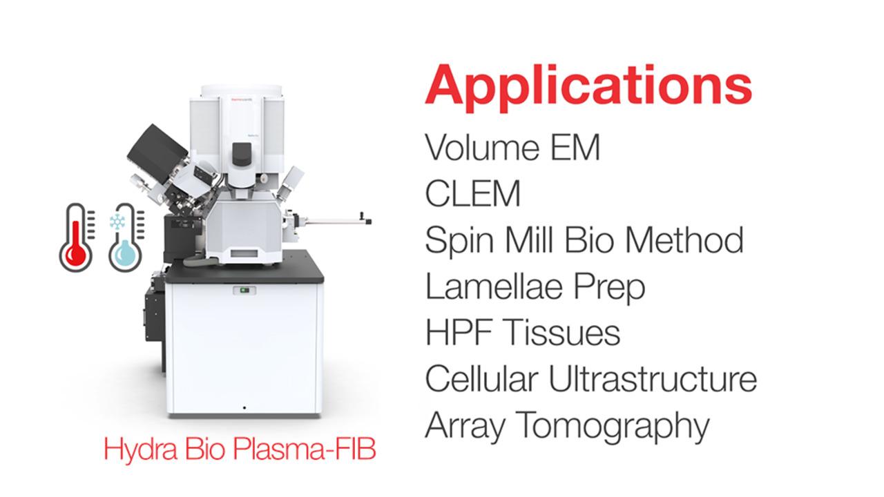
With a state-of-the-art inductively coupled plasma (ICP) focused ion beam and four ion species, the Thermo Scientific Hydra Bio Plasma-FIB helps you find the right ion beam for all your samples. The Hydra Bio PFIB SEM explores tissues to proteins through volume electron microscopy both at cryogenic and room temperatures and cellular cryo-tomography. The four ion species (argon, nitrogen, oxygen, and xenon) are compatible with all commonly used sample-embedding media and protocols like high-pressure tissue freezing. With its advanced software suite, with the Hydra Bio PFIB you can gather consistent, high-quality, gallium-free data from multiple workflows such as high-resolution serial PFIB imaging, millimeter-scale analysis with Spin Mill Bio Method, array tomography, correlative light and electron microscopy and lamellae cryo-lift-out.
Volume imaging for new studies for frozen-hydrated specimens with high-resolution serial PFIB milling and imaging in native state with excellent contrast.
Multiple ion species (Xe, Ar, O, and N) for a range of sample materials and the Thermo Scientific Spin Mill Bio Method, an exclusive technique for large-area planar milling that yields similar sized imaging areas as microtome slicing.
Prepare lamellae from within large (HPF) volumes with plasma FIB, CLEM localization through integrated light and electron imaging with Thermo Scientific iFLM Correlative System and cryo-lift-out with Thermo Scientific Easylift Cryo Lift-Out System.

The Hydra Bio PFIB Cryo Package includes a fully integrated 360° rotation cryo-stage to maintain sample temperature below –170°C and a cryo-sample loading system to ensure that samples remain at cryo temperatures during transfer in and out of the instrument. With the excellent contrast of the Hydra Bio SEM column, uncover sub-cellular details on high pressure frozen and plunge frozen samples. No staining is required. Cryo-fixation avoids the artifacts that are usually associated with classic sample preparation steps with excellent contrast and minimal charging artifacts. Integrated Micro-Sputter Coater reduces charging under cryogenic conditions. Dedicated cryo-lamella preparation software supports your workflow with Thermo Scientific AutoTEM Cryo Software or cryo-volume imaging with Thermo Scientific Auto Slice & View Software.

The Hydra Bio Plasma-FIB features a unique multiple ion species PFIB column with four ion species (Xe, Ar, O, N) with a patented, automated, and fast switching capability (<10 minutes). For example, O+ ion-plasma provides superior data acquisition efficiency and image quality for samples embedded in either epoxy- or acrylic-based resins. Unlike Ga-based FIBs, curtain-free surfaces are easily generated for a wide range of materials such as LR-White, HM20, EPON and Quetol resins.

The unique Thermo Scientific Spin Mill Bio Method on the Hydra Bio Plasma-FIB provides large-area planar milling up to 1 mm in diameter, with a geometry like microtome-based serial block face imaging but at a slice thickness as small as 5 nm. Uncover and visualize large sample areas with Spin Mill Bio Method, then focus in on your regions of interest. It is also ideal to prepare clean and smooth surfaces. The Spin Mill Bio Method is fully automated and easy to set up using guided software. Multiple areas for image acquisition within one experiment can be selected. Each area of interest can be imaged at different imaging settings based on the specificity of the experiment. Sparse features can easily be identified, and statistically relevant 3D data can be collected from multiple areas.

The Easylift Cryo Lift-Out System supports “on the grid” and bulk high pressure frozen (HPF) specimen techniques to help confirm region of interest in final lamella. Fluorescently labeled regions of interested are carefully localized using the iFLM Correlative System, and then extracted and thinned before transferring for tomographic data collection in a cryo-TEM.

The integrated iFLM Correlative System combines fluorescence and electron imaging with ion milling and guided lamella production and supports correlative (CLEM) workflows. Identify, target, and confirm regions of interest without the hassle of transferring between instruments. A suite of shuttles is available, including a Leica External CLEM shuttle, to support grid or common HPF specimen formats. The iFLM Correlative System has been optimized for low distortions across a large field of view and exceptionally low mismatch between color channels. For external data sources Thermo Scientific Maps Software is available for correlation.
For Research Use Only. Not for use in diagnostic procedures.