Search Thermo Fisher Scientific
- Order Status
- Quick Order
-
Don't have an account ? Create Account
Search Thermo Fisher Scientific

The award-winning Thermo Scientific Krios G4 Cryo-TEM is a 300 kV cryo-transmission electron microscope engineered to enable 3D visualization of proteins and molecular machines, their localization, and dynamics within the architecture of the biological cell for unmatched biological insights. See the examples below of how biological scientists have sought to unlock the mysteries of life -- aging, curing disease, and enhancing health using the Krios Cryo-TEM.
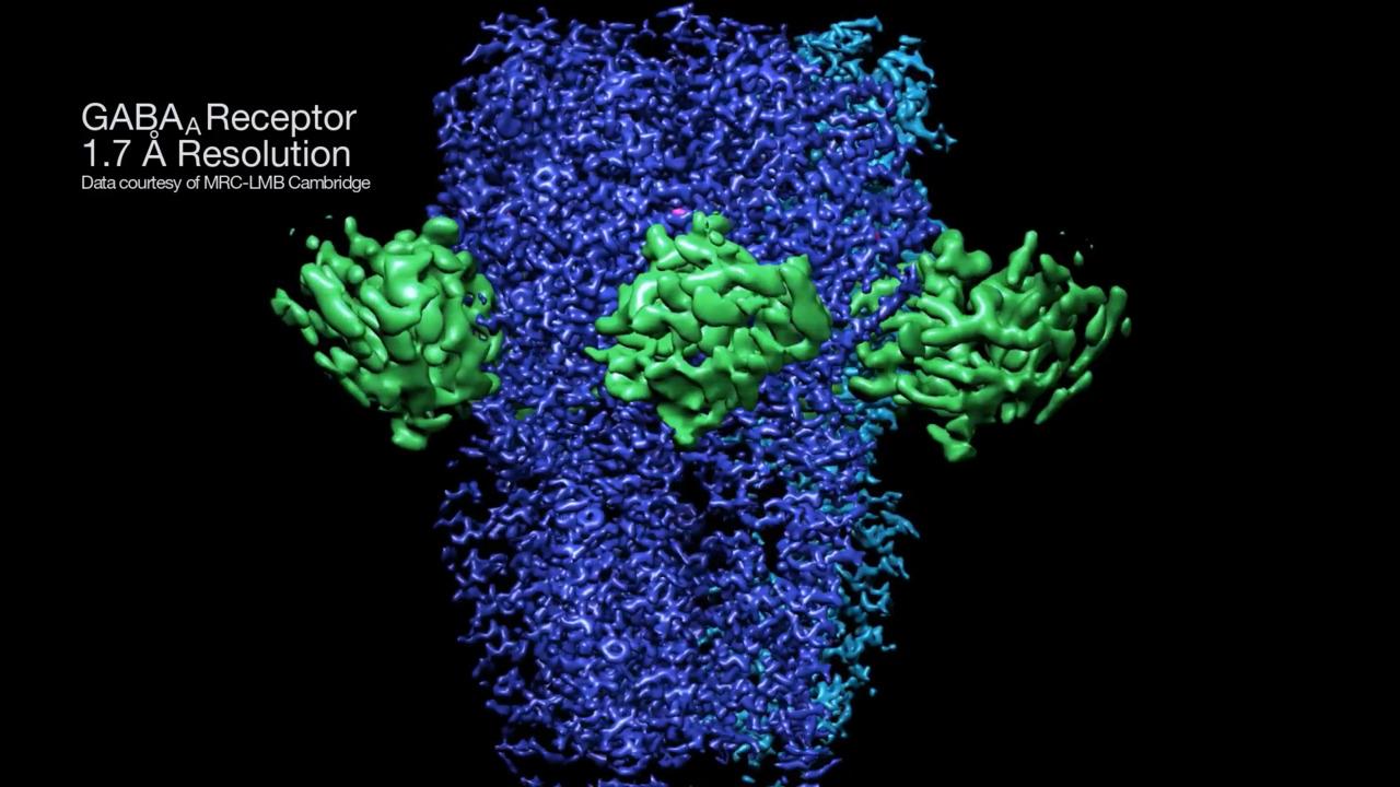
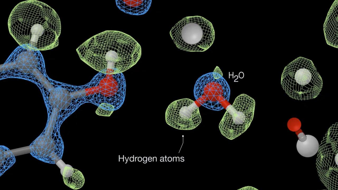
Using a Krios G4 Cryo-TEM equipped with a Thermo Scientific Selectris X Imaging Filter, structural biologists achieved record-breaking resolution results, featured on the cover of Nature and other recent articles from Science and Nature. Scientists from the Medical Research Council Laboratory of Molecular Biology in Cambridge, UK, obtained a 1.2 ångström resolution structure of the iron-storing protein apoferritin. They also achieved a 1.7 ångström resolution of membrane protein (GABA receptor), with an even sharper resolution in key parts of the protein. This human membrane protein has target sites for general anesthetics, benzodiazepines, barbiturates, and neuroactive steroids, making it very relevant to structure-based drug discovery. In addition to the resolution leap, the Selectris technology can also significantly impact productivity. By having a better filter and camera, you can achieve a specific resolution with much less data.
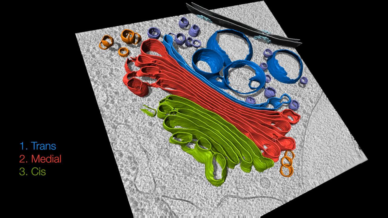
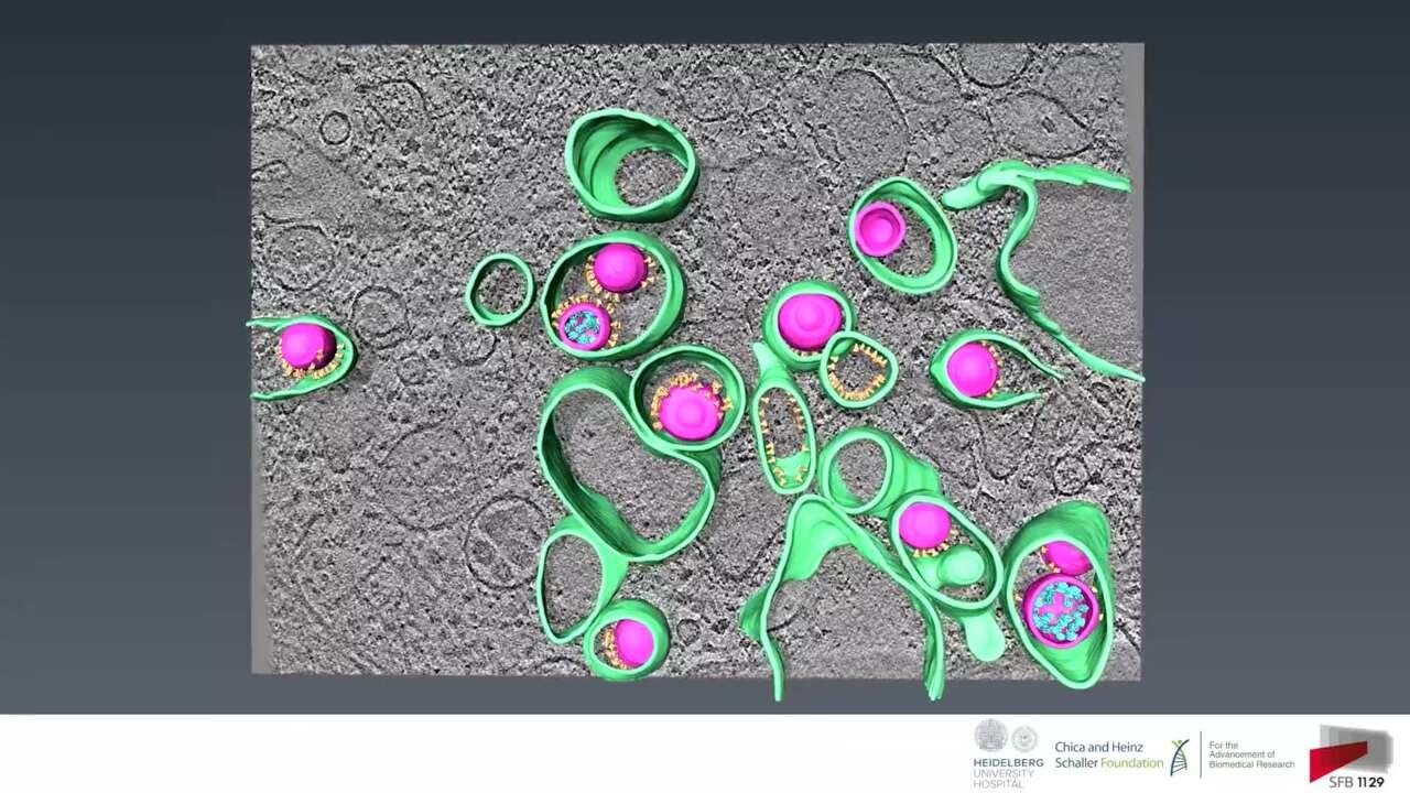
The Krios G4 Cryo-TEM offers best in class cryo-electron tomography (cryo-ET) capabilities, providing nanometer-scale imaging of a cell’s interior in 3D and the visualization of protein complexes within their physiological environments. Using the Krios Cryo-TEM, tomography data can be obtained from whole bacterial cells or lamellae prepared with Thermo Scientific Aquilos, Arctis or Hydra Bio focused ion beam (Cryo-FIB) instruments.
For Research Use Only. Not for use in diagnostic procedures.