Search Thermo Fisher Scientific
- Order Status
- Quick Order
-
Don't have an account ? Create Account
Search Thermo Fisher Scientific

Catalog numbers: HCSDCX7LZRPRO
The Thermo Scientific CellInsight CX7 LZR Pro platform seamlessly integrates advanced screening technologies to deliver superior performance for lightning-fast discovery across the life sciences. Designed to meet the challenging needs of emerging life science models, the CX7 LZR Pro incorporates both laser-based fluorescence illumination and integrated Nipkow spinning-disc confocal technology for publication quality imaging of a broad range of samples from simple monolayers to thick spheroid samples.
Dual mode software- and laser-based autofocusing methods contribute to the platform’s diverse capabilities to investigate simple to complex models. On-the-fly phenotyping enables parallel image capture and analysis for multiplexed cytometry measurements in real time. The Thermo Scientific Amira software option enables 3-dimensional analysis of the most complex research models from 3D morphological tracing in neurons to co-localizations of immune and cancer cells within tumors to enable next-generation translational research.
Browse the features, specifications, videos, sample data, and reagent selection guide sections below.
In addition to the common features of all high-content screening platforms, the CellInsight CX7 LZR Pro platform offers:
Next-generation sCMOS camera
The CX7 LZR Pro platform has been upgraded with a sCMOS camera, offering greater than 95% peak quantum efficiency and low background 1.0e-ready noise. This quantum efficiency further accelerates high-throughput performance thanks to reduced exposure times across all wavelengths. Boost your experiment’s assay window, with up to 65,536 shades of grey detection while not sacrificing background noise thanks to the -20˚C cooling.
Introducing Olympus X-line objectives to the CX7 Pro
The CX7 LZR Pro platform now offers Olympus X-line objectives to further improve the instrument's imaging quality and assay performance. Publication-quality imaging is now standard using the new CX7 LZR Pro platform.
Laser-based illumination
Seven independent lasers aligned with multiplexable fluorescent cellular labeling for the best performance in speed and image quality.
Laser illumination provides flatter and brighter images with low background.
Laser light engine
Illuminates the field evenly resulting in the best image quality appropriate for quantitation.
Up to 6X increase in 3D confocal screening times compared to LED illumination.
Short exposure times and laser autofocus capabilities
Speeds up the acquisition of images.
Near-IR (785 nm) laser excitation
Expand your multiplexing capabilities with near-IR (785 nm) laser excitation.
Autofocus
Dual Mode Autofocusing Software and Laser Autofocus modes accommodate the widest variety of sample types.
Extremely bright illumination
Penetrate thick samples during widefield or confocal 3D imaging.
| Category | Attribute | Description |
| Optics | Camera | Photometrics high-resolution fluorescent camera:
|
| Light source | High-output, laser-based illumination. Configured for imaging in the UV range through near-IR range with the provided filter sets. | |
| Objectives | Standard (Olympus™ objectives):
Optional (Olympus objectives):
|
|
| Image compatibility | JPG, BMP, GIF, PNG, TIF, C01, DIB | |
| Physical characteristics | Dimensions | 20 in. x 32 in. x 18 in. (50.8 cm x 81.3 cm x 45.7 cm) |
| Weight | 68 kg (150 lb) | |
| System | Data management | Compatible with Thermo Scientific Store Image and Database Management Software |
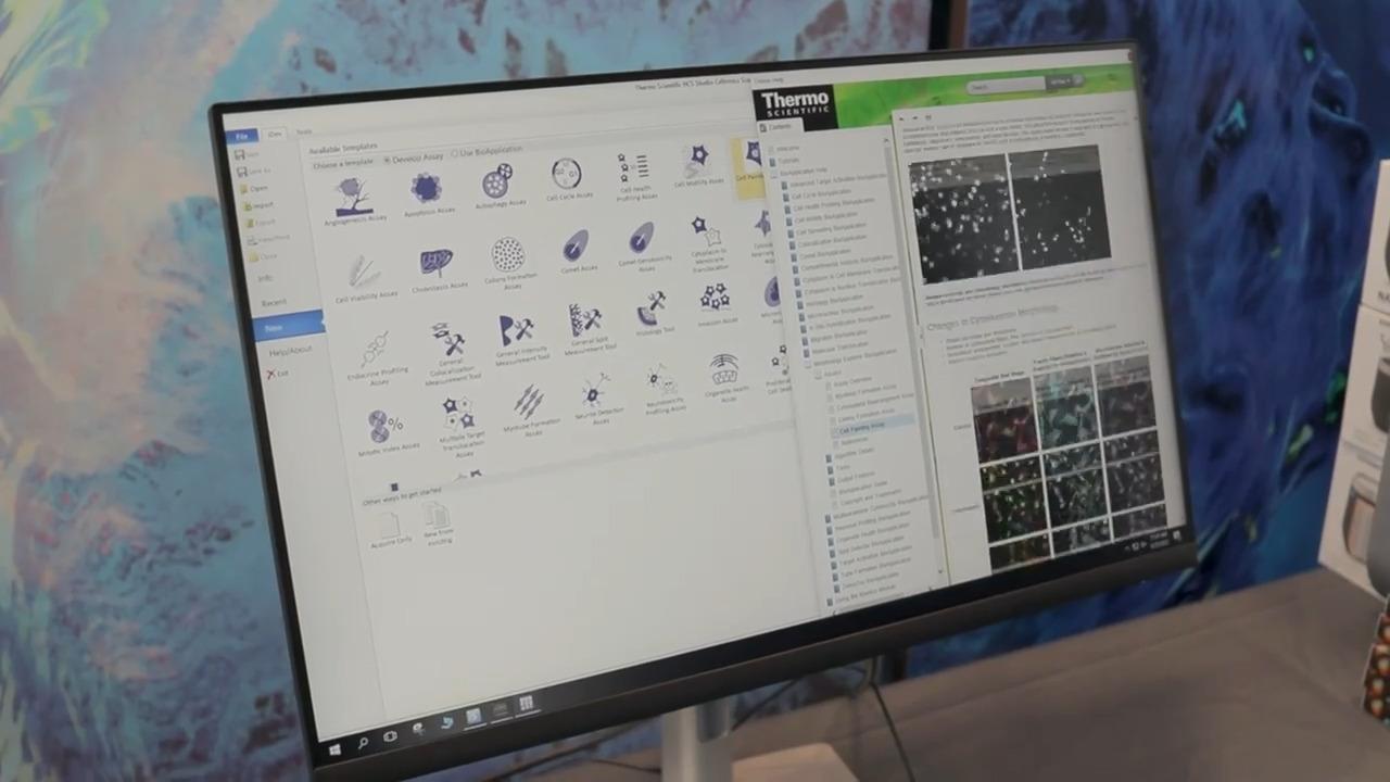
| Software | Thermo Scientific HCS Studio 5.0 Cell Analysis Software with the cell painting bioapplication | |
| PC | Windows 10 Professional 64-Bit PC with 32GB (4X8GB) RAM, Intel® Xeon® Processor 3.7GHz Turbo, RAID I Controller with 1.8TB Hard Drive and 256 Boot Drive. Includes Dell 24" High Resolution Widescreen Monitor or equivalent, keyboard and mouse |
|
| Wattage | 300 W |
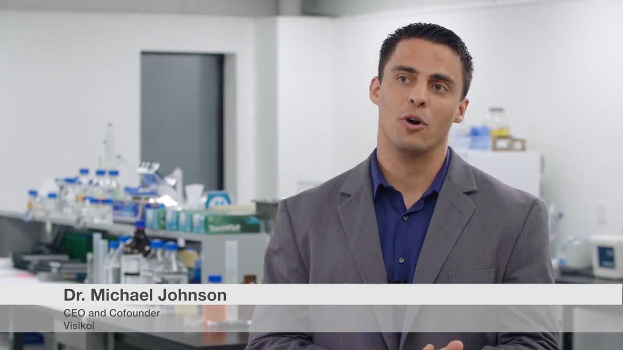
Introducing the Thermo Scientific CellInsight CX7 LZR Pro High-Content Analysis Platform


3D confocal scan of spheroids is faster than LED illumination
3D, 4-color stacks were acquired on A549 spheroids using CellInsight Cx7 LZR with a 10x/0.4 NA objective. 70 µm confocal was used for all 4 colors (20 slices, 7 µm steps). Illumination was via 405, 488, 651, and 647 lasers. The scan improvement speed when using the CellInsight CX7 LZR platform over the LED instrument was 5.8x for a 1-well scan and 6.1x for a 39-well scan.
The Thermo Scientific CellInsight CX7 LZR Pro high-content instrument allows you to take advantage of the entire fluorescence spectrum to optimize your assay—and multiplex your components to ask more in-depth biological questions. Learn more about reagents for cell viability, proliferation, and function or reagents to determine cell structure. For more specific applications using assays and high-content platforms, check out Assay Selection.
For Research Use Only. Not for use in diagnostic procedures.

.png "Immunofluorescence image of HeLa spheroid")

