Search Thermo Fisher Scientific
Cryo-electron tomography workflow
The Thermo Scientific cryo-electron tomography (cryo-ET) workflow is a complete end-to-end workflow covering cell culture to flash freezing cells to final 3D visualization and analysis. In the Thermo Scientific Arctis Cryo-Plasma-FIB, specially designed TomoGrids function to ensure correct lamella alignment to the tomographic tilt axis, from initial milling through high-resolution TEM imaging. The direct connection to any Autoloader-equipped cryo-TEM (e.g.,Thermo Scientific Krios or Thermo Scientific Glacios Cryo-TEMs) eliminates manual grid handling and transfer steps between FIB-SEM and TEM. Combined with Thermo Scientific Amira Software, you can visualize, analyze and obtain quantitative information from your images.
Cryo-FIB sample preparation
Thermo Fisher Scientific is the industry leader in focused ion beam scanning electron microscopy (FIB-SEM) with 30 years of experience as part of our Thermo Scientific DualBeam product line. We offer a broad product portfolio and advanced automation capabilities for a range of applications, including transmission electron microscopy sample preparation as well as subsurface and 3D characterization. The addition of cryo-FIB technology has allowed the integrity of flash-frozen (vitrified) samples to be maintained for a variety of biological and life science applications.
Cryo-transmission electron microscopes for cryo-ET
Heat decontamination for cryo-TEM
Our solution for 60°C heat decontamination allows installation of the Thermo Scientific Krios Cryo-Transmission Electron Microscope (Cryo-TEM) in higher biosafety-level containment facilities (e.g. BSL-3). For these installations, we offer a 60°C heat decontamination solution consisting of both hardware and software to heat the interior of the microscope enclosure.
Cryo-electron tomography software
Tomography 5 and Tomo Live Software
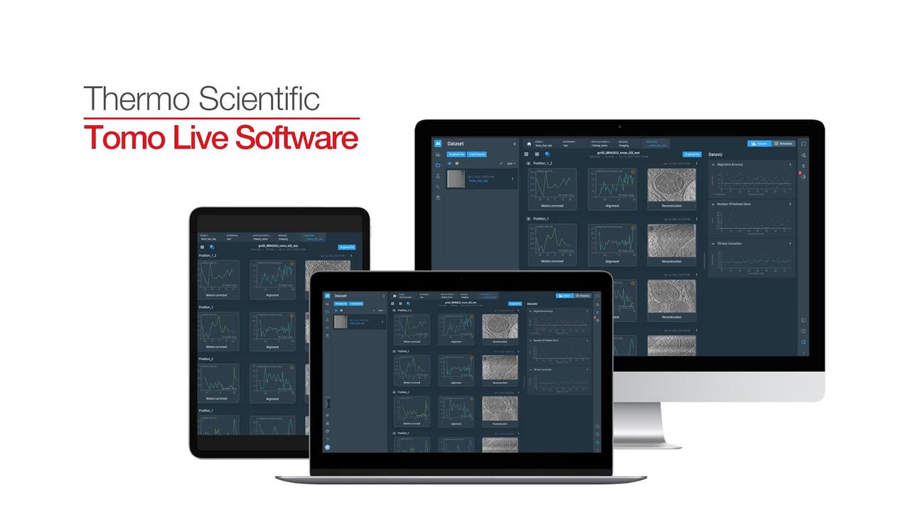
User-friendly batch acquisition software for cryo-electron tomography. Thermo Scientific Tomo Live Software is an optional add-on for Thermo Scientific Tomography 5 Software that enables on-the-fly quality monitoring of data generated with Tomography 5 Software by performing real-time reconstruction of tilt series into 3D volumes.
For Research Use Only. Not for use in diagnostic procedures.
