Search Thermo Fisher Scientific
Simple Automated Imaging Information

Simple automated imaging can be carried out during different stages of the experimental workflow, depending on your need. This could involve hands-off microplate scanning, robotic plate loading or automatic cell counting. This section offers educational tools including application notes, videos, webinars and articles that can help address your specific experimental needs.
Simple automated imaging features
Application note

Cell division and migration during wound healing visualized on the EVOS FL Auto Imaging System
The EVOS FL Auto Imaging System is used with Gibco primary cells and media, Invitrogen detection reagent, and the EVOS Onstage Incubator to observe cell division and migration of neonatal human dermal fibroblast (HDFn) cells in a wound healing assay over a period of 70 hours.
Instrument how-to video
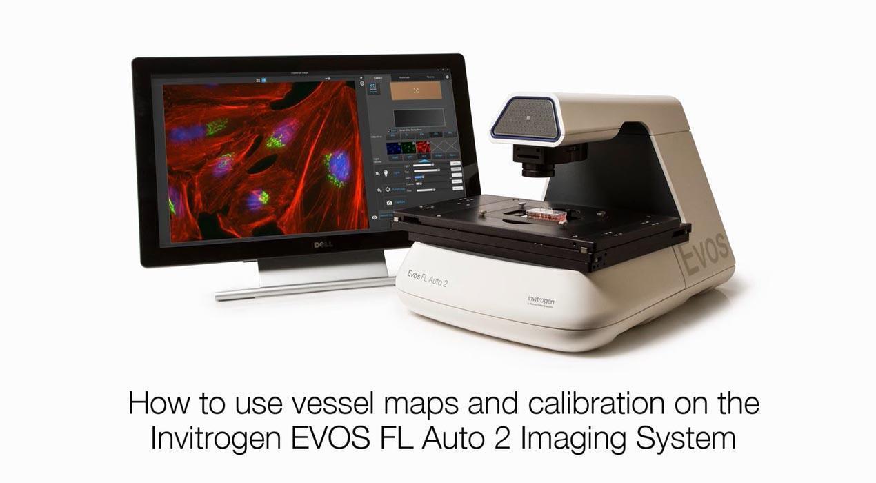
How to setup vessel maps on the EVOS FL Auto 2 microscope
The EVOS FL Auto 2 can accommodate most vessel types and sizes, including slides, multi-well plates, culture flasks, and petri dishes. See how easy it is to setup the EVOS FL Auto 2 for imaging with the vessel maps tool.
Simple automated imaging learning resources
No records were found matching your criteria
| Type | Title | Categories |
|---|---|---|
| Application note (2015) | Using time-lapse imaging with the EVOS FL Auto Imaging System | EVOS FL Auto Microscope, fluorescence microscopy/fluorescence imaging, hypoxia, live-cell imaging, onstage incubator |
| Application note (2015) | Guide to setting up hypoxic conditions on the EVOS FL Auto Imaging System with Onstage Incubator | EVOS FL Auto Microscope, fluorescence microscopy/fluorescence imaging, live-cell imaging, onstage incubator, phagocytosis |
| Application note (2015) | Quantitation of proliferating cells with the EVOS FL Auto Imaging System | antibodies, antibody labeling, ArrayScan, cell proliferation, EVOS FL Auto Microscope, fluorescence microscopy/fluorescence imaging, fluorescent dyes, high content analysis, onstage incubator, phagocytosis |
| Application note (2015) | Phagocytosis visualized on the EVOS FL Auto Imaging System with Onstage Incubator | CellLight, EVOS, fluorescence microscopy/fluorescence imaging, live-cell imaging, ReadyProbes |
| Application note (2015) | Image tiling and stitching using the EVOS FL Auto Imaging System | cell proliferation, EVOS FL Auto Microscope, fluorescence microscopy/fluorescence imaging, Imaging sample preparation, ReadyProbes |
| Application note (2015) | Fluorescent viability assays on the Countess II FL Automated Cell Counter | automated cell counter, cell counting, Countess, EVOS, fluorescent dyes, ReadyProbes, viability |
| Application note (2015) | Fluorescent protein reporter gene transduction efficiency measured with the Countess II FL Automated Cell Counter | automated cell counter, cell counting, Countess, EVOS, fluorescent dyes, genetic transduction |
| Application note (2015) | Fluorescent apoptosis evaluation on the Countess II FL Automated Cell Counter | apoptosis, automated cell counter, cell counting, Countess, EVOS, fluorescent dyes, viability |
| Application note (2015) | Collecting Z-stack image sequences with the EVOS FL Auto Imaging System | cell structure-all, CellLight, EVOS FL Auto Microscope, fluorescence microscopy/fluorescence imaging, live-cell imaging, ReadyProbes |
| Application note (2015) | Cell division and migration during wound healing visualized on the EVOS FL Auto Imaging System | cell health, cell proliferation, EVOS FL Auto Microscope, fluorescence microscopy/fluorescence imaging |
| Application note (2016) | Blood cell counting using the Countess II FL Automated Cell Counter | automated cell counter, brightfield microscopy, cell counting, cell counting sample preparation, Countess, EVOS, fluorescent dyes, immunophenotyping, ReadyProbes |
| BioProbes article (2013) | The EVOS FL Auto Imaging System—Automated imaging. Simplified | brightfield microscopy, cell structure-all, EVOS, fluorescence microscopy/fluorescence imaging |
| BioProbes article (2014) | Introducing a three-channel cell counter for your benchtop—The new and improved Countess II FL Automated Cell Counter | cell counting, automated cell counter, brightfield microscopy, Countess, EVOS fluorescence microscopy/fluorescence imaging |
| BioProbes article (2015) | Fluorescence-based viability assays for the Countess II FL Automated Cell Counter—Incorporate automation into your cell analysis workflows | automated cell counter, cell counting, Countess, EVOS, fluorescence microscopy/fluorescence imaging, viability |
| Tutorial | 5.3 Image capture with EVOS FL Auto 2.0–Fixed cell imaging: 5 steps for publication-quality images | EVOS FL Auto 2.0, fixed-cell imaging, fluorescence microscopy/fluorescence imaging, image capture, immunofluorescence (IF) |
| Video | Setting up an automated scan routine on the EVOS FL Auto 2 Microscope Save time when imaging cells by easily setting up an automated scan routine using the EVOS FL Auto 2 microscope. | EVOS, fluorescence microscopy/fluorescence imaging, instrument set-up |
| Video | Adding oil to objectives on the EVOS FL Auto 2 Microscope See how easy it is to add oil to your objective without having to remove your sample when imaging with the EVOS FL Auto 2 Microscope. | EVOS FL Auto 2 Microscope, fluorescence microscopy/fluorescence imaging, instrument set-up |
| Video | Reviewing Cell Images on the EVOS FL Auto 2 Microscope Cell images can be saved from the EVOS FL Auto 2 as a single field, scan, time lapse, and z-stack. Easily review your images and metadata or make adjustments to image brightness, contrast, or gamma and resave your images with the new settings. | EVOS FL Auto 2 Microscope, fluorescence microscopy/fluorescence imaging, instrument set-up |
| Video | Using the locations tool on the EVOS FL Auto 2 Microscope Learn how to create custom locations to use in automated routines for scanning and time lapse experiments on the EVOS FL Auto 2 Cell Imaging System. | EVOS, fluorescence microscopy/fluorescence imaging, instrument set-up |
| Video | How to setup vessel maps on the EVOS FL Auto 2 Microscope The EVOS FL Auto 2 can accommodate most vessel types and sizes, including slides, multi-well plates, culture flasks, and petri dishes. See how easy it is to setup the EVOS FL Auto 2 for imaging with the vessel maps tool. | EVOS FL Auto 2 Microscope, fluorescence microscopy/fluorescence imaging, instrument set-up |
| Video | How to add a scale bar and grid to cell images on the EVOS FL Auto 2 Microscope See how easy it is to add a scale bar and grid to your live or captured cell images on the EVOS FL Auto Microscope. You can adjust the grid size, color, and alignment to fit the needs of your experiment. | EVOS FL Auto 2 Microscope, fluorescence microscopy/fluorescence imaging, instrument set-up |
| Video | Create z-stacks using the EVOS FL Auto 2 Microscope Capturing images at different Z-planes can be a powerful tool in fluorescence microscopy. This capability can reveal conditions not seen with standard widefield imaging and easier than ever to use with the EVOS FL Auto 2 microscope. | EVOS FL Auto 2 Microscope, fluorescence microscopy/fluorescence imaging, instrument set-up |
| Video | Z Stack image of HeLa cells labeled with CellLights reagents A series of images were captured on the EVOS FL Auto Cell Imaging System. Creating a Z-stack from these images allowed the observation of cellular cytoskeletal changes, which can be indicative of the loss of cell health. Methods HeLa cells grown in MatTek 6-well glass bottom culture plates were transduced with CellLights Tubulin-GFP and CellLight Mitochondria-RFP overnight at 37oC. The following day, NucBlue Live reagent (2 drops/mL) was added to the cultures. Cells were then imaged on an EVOS FL Auto Cell Imaging System with 100x oil immersion objective using the Z-stack function. The step size was set using the Nyquist formula and performed at 0.366 μm. | cell health, cell structure-all, CellLight, EVOS FL Auto Microscope, fluorescence microscopy/fluorescence imaging, fluorescent proteins, live-cell imaging |
| Video | Time-lapse of cell migration and apoptosis during angiogenesis Angiogenesis pseudo-tube formation and apoptosis shown with time-lapse imaging. The time-lapse images were captured and assembled using an EVOS FL Auto Imaging System equipped with the EVOS Onstage Incubator. | angiogenesis, apoptosis, EVOS FL Auto Microscope, fluorescence microscopy/fluorescence imaging, live-cell imaging, onstage incubator |
| Webinar | Fixed cell imaging—Five steps for publication-quality images With over 40 years dedicated to cell imaging research, we offer long-proven tools and protocols to help confidently create quality cell images the first time. This on demand webinar covers the 5 essential steps to getting great images. | antibodies, blocking, Celleste, dyes, EVOS FL Auto 2.0, fixation, fixed-cell imaging, fluorescence microscopy/fluorescence imaging, immunofluorescence (IF), sample detection, sample labeling, sample preparation, signal amplification |
| White paper (2015) | Accuracy and precision with the Countess II FL Automated Cell Counter | automated cell counter, brightfield microscopy, cell counting, Countess |
For Research Use Only. Not for use in diagnostic procedures.