Search Thermo Fisher Scientific
Cell Viability, Proliferation and Cell Cycle Information

Measuring cell health parameters like cell viability, cell proliferation and cell cycle are critical to understanding and interpreting your results. Here you will find educational resources such as application notes, webinars, videos, articles and more that cover the use of many of our reagents and kits for monitoring cell function.
Cell viability, proliferation and cell cycle features
BioProbes article

Multiplex assays for robust cell health analyses
Plate-based viability assays are a fundamental tool in drug discovery for evaluating the potency of compounds and the sensitivity of cell lines to specific agents
Technology video
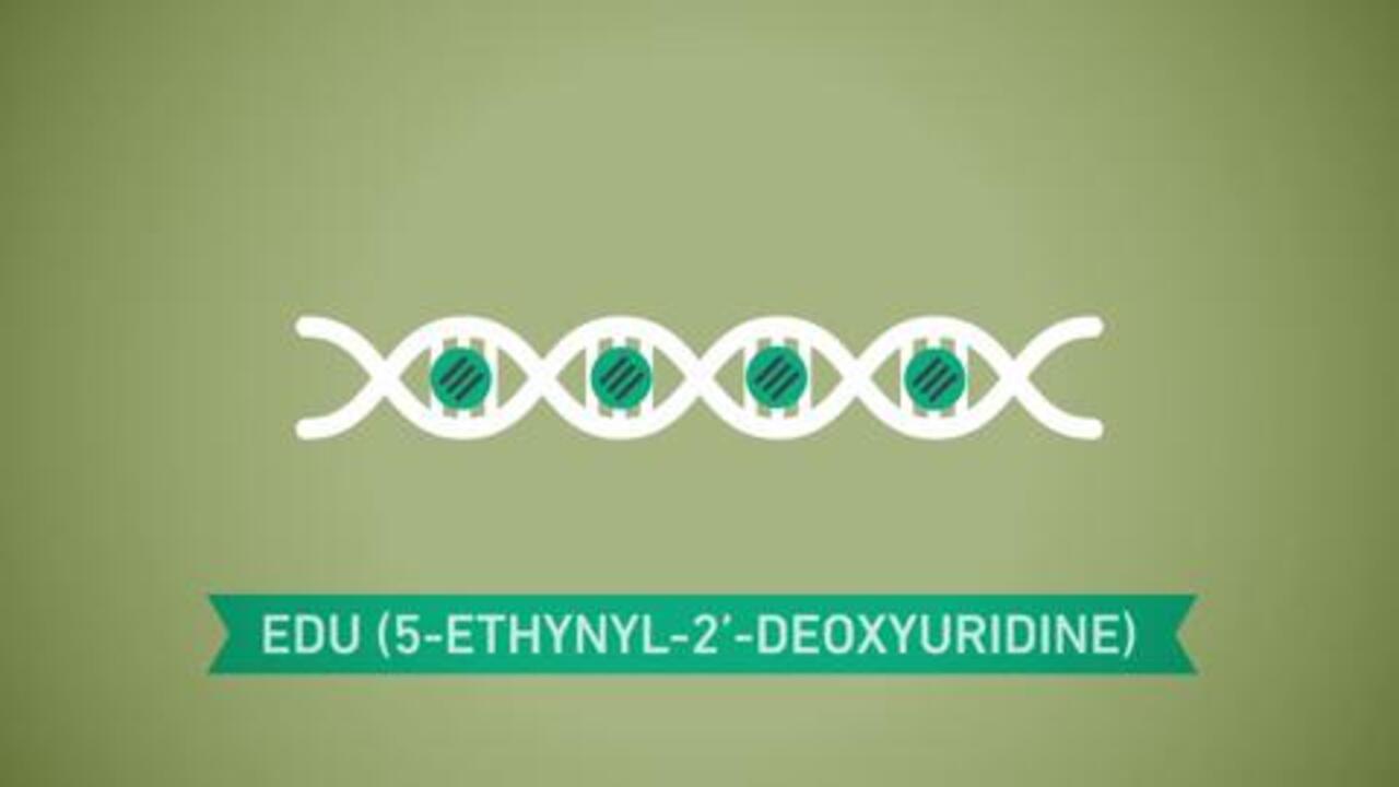
How the Click-iT Plus EdU Proliferation Assay works
Measuring DNA synthesis is the most precise way to detect changes in cell proliferation. Image-based proliferation assays generate spatial and temporal results that cannot be detected with other methods. This video describes and compares two of the most referenced image-based proliferation technologies.
Cell viability, proliferation and cell cycle learning resources
No records were found matching your criteria
| Type | Title | Categories |
|---|---|---|
| Application note (2011) | Bacterial analysis using the Attune Acoustic Focusing Cytometer | Attune/Attune NxT, flow cytometer/flow cytometry, fluorescent dyes, microbiology, viability |
| Application note (2011) | Detection of human mesenchymal stem cells using the Attune Acoustic Focusing Cytometer | Attune/Attune NxT, cell cycle, flow cytometer/flow cytometry, immunophenotyping, stem cell research |
| Application note (2012) | Monitoring neurite morphology and synapse formation in primary neurons for neurotoxicity assessments and drug screening | ArrayScan, drug discovery, fluorescence microscopy/fluorescence imaging, fluorescent dyes, high content analysis, microplate reader, neuroscience, viability |
| Application note (2012) | Microbiological applications using the Attune Acoustic Focusing Cytometer | Attune/Attune NxT, cell health, flow cytometer/flow cytometry, fluorescent dyes, microbiology, viability |
| Application note (2013) | Multiplexed mitosis and apoptosis analysis | antibodies, apoptosis, ArrayScan, cell cycle, cell proliferation, fluorescence microscopy/fluorescence imaging, fluorescent dyes, high content analysis, microplate reader, particles |
| Application note (2014) | High content cell cycle screening: Pairing FUCCI technology with Thermo Scientific HCS Studio 2.0 Software and FCS Express Image cytometry | ArrayScan, cancer, cell cycle, drug discovery, fluorescence microscopy/fluorescence imaging, fluorescent dyes, high content analysis, microplate reader |
| Application note (2015) | Quantitation of proliferating cells with the EVOS FL Auto Imaging System | antibodies, antibody labeling, ArrayScan, cell proliferation, EVOS FL Auto Microscope, fluorescence microscopy/fluorescence imaging, fluorescent dyes, high content analysis, onstage incubator, phagocytosis |
| Application note (2015) | Image tiling and stitching using the EVOS FL Auto Imaging System | cell proliferation, EVOS FL Auto Microscope, fluorescence microscopy/fluorescence imaging, Imaging sample preparation, ReadyProbes |
| Application note (2015) | Fluorescent viability assays on the Countess II FL Automated Cell Counter | automated cell counter, cell counting, Countess, EVOS, fluorescent dyes, ReadyProbes, viability |
| Application note (2015) | Fluorescent apoptosis evaluation on the Countess II FL Automated Cell Counter | apoptosis, automated cell counter, cell counting, Countess, EVOS, fluorescent dyes, viability |
| Application note (2015) | Cell division and migration during wound healing visualized on the EVOS FL Auto Imaging System | cell health, cell proliferation, EVOS FL Auto Microscope, fluorescence microscopy/fluorescence imaging |
| Application note (2015) | Live/dead algal analysis with the Attune NxT Flow Cytometer | algae, Attune/Attune NxT, flow cytometer/flow cytometry, fluorescent dyes, viability |
| BioProbes articles (Issues 50– present day) | BioProbes Journal of Cell Biology Application | cell analysis, flow cytometry, imaging microscopy, immunoassays, antibodies, protein detection and quantification |
| Molecular Probes Handbook | Nucleic acid stains—Section 8.1 | cell cycle, cell structure-nucleus, cell structure-plasma membrane, cell viability, DAPI, DNA binding dyes, flow cytometer/flow cytometry, fluorescence microscopy/fluorescence imaging, fluorescent dyes, fluorometer, membrane permeability, RNA binding dyes, SYTO, SYTOX |
| Molecular Probes Handbook | Viability and cytotoxicity assay reagents—Section 15.2 | cell health, cell viability and function, flow cytometer/flow cytometry, fluorescence microscopy/fluorescence imaging, microplate reader |
| Molecular Probes Handbook | Viability and cytotoxicity assay kits for diverse cell types—Section 15.3 | cell health, cell viability and function, flow cytometer/flow cytometry, fluorescence microscopy/fluorescence imaging, microplate reader |
| Molecular Probes Handbook | Overview of probes for cell viability, cell proliferation and live-cell function—Section 15.1 | cell health, cell death, cell proliferation, cell viability and function |
| Molecular Probes Handbook | Assays for cell enumeration, cell proliferation and cell cycle—Section 15.4 | cell counting, cell cycle, cell proliferation |
| Protocol | Vybrant MTT Cell Proliferation Assay Kit | cell viability, microplate |
| Protocol | Viability staining protocol for flow cytometry | cell viability, flow cytometry |
| Protocol | SYTOX Dead Cell Stains | cell viability, flow cytometry |
| Protocol | Red blood cell lysis protocols using eBioscience lysis buffers | cell proliferation, flow cytometry |
| Protocol | ReadyProbes Cell Viability Imaging Kit, Blue/Red | cell viability, imaging |
| Protocol | ReadyProbes Cell Viability Imaging Kit, Blue/Green | cell viability, imaging |
| Protocol | PrestoBlue Cell Viability Reagent for microplates | cell viability, microplate |
| Protocol | PrestoBlue and CyQUANT Direct Confirmation Assay | cell viability, microplate |
| Protocol | Pharmacological induction of apoptosis with camptothecin | cell vitality, flow cytometry, high content analysis, imaging, microplate |
| Protocol | NucRed Dead 647 ReadyProbes Reagent for viability | cell viability, imaging |
| Protocol | NucGreen Dead 488 ReadyProbes Reagent for viability | cell viability, imaging |
| Protocol | LIVE/DEAD Yeast Viability Kit | cell viability, imaging |
| Protocol | LIVE/DEAD Violet Viability/Vitality Kit | cell viability and cell vitality, flow cytometry |
| Protocol | LIVE/DEAD Viability/Cytotoxicity Kit for mammalian cells | cell viability, imaging |
| Protocol | LIVE/DEAD Sperm Viability Kit for flow cytometry | cell viability, flow cytometry |
| Protocol | LIVE/DEAD Sperm Viability Kit for imaging | cell viability, imaging |
| Protocol | LIVE/DEAD Fixable Dead Cell Stains | cell viability, flow cytometry |
| Protocol | LIVE/DEAD Cell Imaging Kit (488/570) | cell viability, imaging |
| Protocol | LIVE/DEAD BacLight Bacterial Viability Kit | cell viability, imaging |
| Protocol | LIVE BacLight Bacterial Gram Stain Kit | cell viability, imaging |
| Protocol | Image-iT DEAD Green Kit | cell viability, imaging |
| Protocol | HCS Mitochondrial Health Kit | cell viability, high content analysis |
| Protocol | HCS LIVE/DEAD Green Kit using Hoechst 33342 | cell viability, high content analysis |
| Protocol | HCS LIVE/DEAD Green Kit using HCS NuclearMask Deep Red | cell viability, high content analysis |
| Protocol | Staining cells with eFluor proliferation dyes for flow cytometry | cell proliferation, flow cytometry |
| Protocol | CyQUANT Direct Microplate Reagent for cell viability | cell viability, microplate |
| Protocol | Click-iT Plus EdU imaging kits | cell proliferation, imaging |
| Protocol | Click-iT EdU imaging kits | cell proliferation, imaging |
| Protocol | Click-iT EdU HCS assays | cell proliferation, imaging |
| Protocol | CellTrace Violet Cell Proliferation Kit | cell proliferation, flow cytometry |
| Protocol | CellTrace Far Red Cell Proliferation Kit | cell proliferation, flow cytometry |
| Protocol | CellTrace CFSE Cell Proliferation Kit | cell proliferation, flow cytometry |
| Protocol | Cell preparation for flow cytometry protocols | cell proliferation, flow cytometry |
| Protocol | BrdU staining protocol for flow cytometry | cell proliferation, flow cytometry |
| Protocol | BrdU labeling and detection | cell proliferation, imaging |
| Protocol | Annexin V staining protocol for flow cytometry | cell vitality, flow cytometry |
| Scientific poster (2006) | LipidTOX dyes for adipocyte staining in routine imaging applications | cell differentiation, fluorescence microscopy/fluorescence imaging, fluorescent dyes, high content analysis, lipid detection, lipids, stem cell research, viability |
| Scientific poster (2006) | Cell cycle analysis using microplate cytometry: A comparison of laser and dye combinations | Acumen Explorer, cell cycle, fluorescence microscopy/fluorescence imaging, fluorescent dyes, high content analysis, microplate reader |
| Scientific poster (2006) | New fluorescent reagents for the imaging-based assays for the analysis of drug-induced perturbations of cellular lipid metabolism | ArrayScan, fluorescence microscopy/fluorescence imaging, fluorescent dyes, high content analysis, lipid detection, lipids, microplate reader, viability |
| Scientific poster (2006) | Utility of 405 nm-excitable dyes in high content screening using an Acumen Explorer Microplate Cytometer | Acumen Explorer, apoptosis, cell cycle, cell health, fluorescence microscopy/fluorescence imaging, fluorescent dyes, high content analysis, microplate reader, violet laser-excited reagents |
| Scientific poster (2006) | Novel violet-excited reagents for detection of viability and vitality | Alexa Fluor, cell health, CellTrace, flow cytometer/flow cytometry, fluorescent dyes, viability, violet laser-excited reagents |
| Scientific poster (2007) | Evaluation of click chemistry-based alternative to BrdU antibody labeling in tissue and cultured cells using fluorescence microscopy and flow cytometry | antibodies, ArrayScan, cell cycle, cell proliferation, Click-iT, flow cytometer/flow cytometry, fluorescence microscopy/fluorescence imaging, fluorescent dyes, microplate reader, nucleic acid labeling |
| Scientific poster (2007) | A new approach for the detection of intracellular glutathione by fluorescence microscopy and flow cytometry | ArrayScan, flow cytometer/flow cytometry, fluorescence microscopy/fluorescence imaging, fluorescent dyes, glutathione, live-cell imaging, microplate reader, viability, violet laser-excited reagents |
| Scientific poster (2007) | Click chemistry-based detection of S-phase adherent cells using automated microscopy and image analysis | Alexa Fluor, antibodies, ArrayScan, cell cycle, Click-iT, fluorescence microscopy/fluorescence imaging, fluorescent dyes, high content analysis, microplate reader |
| Scientific poster (2007) | Click catalyzed nucleic acid labeling as a novel replacement for BrdU antibody-based cell proliferation assay | Alexa Fluor, antibodies, antibody labeling, cell cycle, cell proliferation, Click-iT, fluorescence microscopy/fluorescence imaging, fluorescent dyes, high content analysis, nucleic acid labeling |
| Scientific poster (2008) | Characterization of DNA content, cyclin B1 and phosphorylated histone H3 with direct S-phase using EDU incorporation in multiparameter testing of cell lines with cell cycle blocking agents | Alexa Fluor, antibodies, apoptosis, ArrayScan, cell cycle, cell proliferation, Click-iT, flow cytometer/flow cytometry, fluorescence microscopy/fluorescence imaging, fluorescent dyes, high content analysis |
| Scientific poster (2008) | Novel Click-IT TUNEL Assay for detection of cell death | Alexa Fluor, antibodies, apoptosis, Click-iT, cytotoxicity, fluorescence microscopy/fluorescence imaging, fluorescent dyes, gel electrophoresis, high content analysis, immunocytochemistry (ICC), viability |
| Scientific poster (2008) | Effects of the thymidine analogues EdU and BrdU on cell viability and cycle progression | Alexa Fluor, cell cycle, cell proliferation, Click-iT, flow cytometer/flow cytometry, fluorescence microscopy/fluorescence imaging, fluorescent dyes, viability |
| Scientific poster (2008) | Detection of S-phase cell cycle progression using 5-ethynyl-2'-deoxyuridine incorporation with click chemistry | Alexa Fluor, antibodies, ArrayScan, cell cycle, Click-iT, flow cytometer/flow cytometry, fluorescent dyes, high content analysis, microplate reader, nucleic acid labeling, nucleic acid quantitation |
| Scientific poster (2008) | Detection of DNA synthesis by automated microscopy and image analysis: Comparison of BRDU method and a new click chemistry-based EDU method | Alexa Fluor, antibodies, ArrayScan, cell cycle, cell proliferation, Click-iT, flow cytometer/flow cytometry, fluorescent dyes, high content analysis, microplate reader, nucleic acid labeling |
| Scientific poster (2009) | Cell-based assays for predictive hepatotoxicity measurements using high content imaging | Alexa Fluor, ArrayScan, cell structure-plasma membrane, Click-iT, fluorescence microscopy/fluorescence imaging, fluorescent dyes, glutathione, high content analysis, microplate reader, nucleic acid quantitation, viability |
| Scientific poster (2009) | Monitoring mitotic cells and DNA content by automated imaging and analysis | Alexa Fluor, cell cycle, Click-iT, fluorescence microscopy/fluorescence imaging, fluorescent dyes, high content analysis, nucleic acid labeling, nucleic acid quantitation, viability |
| Scientific poster (2009) | Dual pulse labeling of S-phase population using click chemistry to measure changes in cell proliferation | Alexa Fluor, cancer, cell cycle, cell proliferation, Click-iT, flow cytometer/flow cytometry, fluorescent dyes |
| Scientific poster (2009) | Proliferative and phenotypic characterization of human mesenchymal stem cells by flow cytometry | Alexa Fluor, cell proliferation, Click-iT, flow cytometer/flow cytometry, fluorescent dyes, immunophenotyping, nucleic acid quantitation, stem cell research, violet laser-excited reagents |
| Scientific poster (2009) | Dual pulse labeling using a new thymidine analog 5-ethynyl-2'-deoxyuridine (EdU) to detect alterations in S-phase progression by fluorescence microscopy and flow cytometry | Alexa Fluor, cell cycle, cell proliferation, Click-iT, flow cytometer/flow cytometry, fluorescence microscopy/fluorescence imaging, fluorescent dyes |
| Scientific poster (2009) | High content imaging of endocytosis & phagocytosis using pHrodo conjugates | Alexa Fluor, ArrayScan, fluorescence microscopy/fluorescence imaging, fluorescent dyes, fluorescent proteins, high content analysis, pH detection, phagocytosis, viability |
| Scientific poster (2009) | Click chemistry-based detection of nascent RNA synthesis using high content imaging and fluorescence microscopy | Alexa Fluor, ArrayScan, Click-iT, fluorescence microscopy/fluorescence imaging, fluorescent dyes, high content analysis, microplate reader, nucleic acid labeling, viability |
| Scientific poster (2009) | A simple, fast and quantitative single-step dead-cell indicator for flow cytometry | apoptosis, flow cytometer/flow cytometry, fluorescent dyes, multicolor flow cytometry, nucleic acid quantitation, viability |
| Scientific poster (2009) | High content analysis of cytotoxicity by laser scanning fluorescence microplate cytometry | Alexa Fluor, Click-iT, fluorescence microscopy/fluorescence imaging, fluorescent dyes, high content analysis, microplate reader, viability |
| Scientific poster (2009) | Accuracy and precision comparison of the hemocytometer and automated cell counting methods | automated cell counter, cell counting, Countess, hemocytometer, particles, viability |
| Scientific poster (2009) | Dead cell stains in flow cytometry: A comprehensive analysis | apoptosis, flow cytometer/flow cytometry, fluorescent dyes, immunophenotyping, viability |
| Scientific poster (2009) | High content imaging and analysis of mitotoxicity and cytotoxicity in fixed cells | cell structure-mitochondria, fluorescence microscopy/fluorescence imaging, fluorescent dyes, high content analysis, viability |
| Scientific poster (2009) | Quantitative analysis of genotoxicity and cytotoxicity to DNA damaging agents using high-content imaging | Alexa Fluor, antibodies, Click-iT, fluorescence microscopy/fluorescence imaging, fluorescent dyes, high content analysis, nucleic acid labeling, nucleic acid quantitation, viability |
| Scientific poster (2009) | Click chemistry-based assays for fluorescence microscopy and high content imaging | Alexa Fluor, apoptosis, Click-iT, fluorescence microscopy/fluorescence imaging, fluorescent dyes, high content analysis, nucleic acid detection, protein detection, viability |
| Scientific poster (2010) | Acoustic focusing cytometry: Sensitivity and throughput | apoptosis, Attune/Attune NxT, cell cycle, flow cytometer/flow cytometry, fluorescent dyes, immunophenotyping, phagocytosis |
| Scientific poster (2010) | Analysis of proliferation dynamics in primary and immortalized human cells | Alexa Fluor, cancer, cell proliferation, CellTrace, circulatory system, Click-iT, flow cytometer/flow cytometry, fluorescent dyes, fluorescent proteins, nucleic acid quantitation |
| Scientific poster (2010) | Antibody- and fluorescent protein-based approaches to measuring autophagy in mammalian cells by fluorescence microscopy | Alexa Fluor, antibodies, autophagy, BacMam technology, cell proliferation, Click-iT, fluorescence microscopy/fluorescence imaging, fluorescent dyes, high content analysis, live-cell imaging |
| Scientific poster (2010) | Proliferative and phenotypic characterization of human mesenchymal stem cells by flow cytometry and imaging | Alexa Fluor, cell proliferation, Click-iT, flow cytometer/flow cytometry, fluorescence microscopy/fluorescence imaging, fluorescent dyes, immunofluorescence (IF), immunophenotyping, stem cell research, violet laser-excited reagents |
| Scientific poster (2010) | Novel tools to enable high resolution gene expression by investigating the nascent transcriptome | Alexa Fluor, Click-iT, fluorescence microscopy/fluorescence imaging, fluorescent dyes, gene expression, high content analysis, magnetic beads, nucleic acid labeling, PCR/qPCR, viability |
| Scientific poster (2010) | A comparison of three techniques to induce efficient ex vivo T cell expansion | Alexa Fluor, antibodies, cell proliferation, CellTrace, flow cytometer/flow cytometry, fluorescent dyes, immune system, magnetic beads, multicolor flow cytometry, viability |
| Scientific poster (2010) | New fluorescent probes for live-cell imaging | autophagy, cell cycle, CellLight, flow cytometer/flow cytometry, fluorescence microscopy/fluorescence imaging, fluorescent proteins, live-cell imaging, pH detection, phagocytosis |
| Scientific poster (2010) | Functional characterization of a novel fluorescent dye for proliferation analysis | Alexa Fluor, cell proliferation, CellTrace, flow cytometer/flow cytometry, fluorescent dyes, violet laser-excited reagents |
| Scientific poster (2010) | Non-cytotoxic near-IR DNA stain for cell cycle analysis in living cells | cell cycle, flow cytometer/flow cytometry, fluorescence microscopy/fluorescence imaging, fluorescent dyes, fluorescent proteins, live-cell imaging, nucleic acid labeling, viability |
| Scientific poster (2010) | BacMam gene expression for functional and imaging applications | BacMam technology, cell cycle, fluorescence microscopy/fluorescence imaging, fluorescent dyes, fluorescent proteins, gene expression, high content analysis, ion flux/ion channels |
| Scientific poster (2010) | Dual pulse labeling using EdU and BrdU to monitor alterations in cellular proliferation by fluorescence microscopy and flow cytometry | Alexa Fluor, cell cycle, cell proliferation, Click-iT, drug discovery, flow cytometer/flow cytometry, fluorescence microscopy/fluorescence imaging, fluorescent dyes |
| Scientific poster (2011) | Cell-based measurements of oxidative stress with a new near infrared emitting probe for reactive oxygen species: Applications for multiplexed evaluation of cell health | ArrayScan, cell health, flow cytometer/flow cytometry, fluorescence microscopy/fluorescence imaging, fluorescent dyes, high content analysis, microplate reader, oxidative stress, viability |
| Scientific poster (2011) | Primary human cells from Life Technologies: Offerings, scale and applications in HCS and HTS formats | Alexa Fluor, antibodies, autophagy, BacMam technology, cell proliferation, cell signaling, Click-iT, drug discovery, fluorescence microscopy/fluorescence imaging, fluorescent dyes, high content analysis |
| Scientific poster (2011) | Validation of high content imaging and analysis assays for cell health and cytotoxicity | antibodies, apoptosis, ArrayScan, autophagy, caspase substrates, cell health, cell proliferation, CellLight, Click-iT, fluorescence microscopy/fluorescence imaging, fluorescent dyes, high content analysis, microplate reader, pH detection |
| Scientific poster (2011) | The next step in the analysis of lymphocyte proliferation | Alexa Fluor, antibodies, Attune/Attune NxT, cell proliferation, CellTrace, flow cytometer/flow cytometry, fluorescent dyes, immunophenotyping, live-cell imaging, violet laser-excited reagents |
| Scientific poster (2011) | Application of acoustic cytometry in microbiology | Attune/Attune NxT, cell concentration, cell health, flow cytometer/flow cytometry, fluorescent dyes, microbiology, viability |
| Scientific poster (2012) | Rapid, in vitro T lymphocyte cell tracing by acoustic focusing cytometry | Attune/Attune NxT, cell proliferation, CellTrace, flow cytometer/flow cytometry, fluorescent dyes, magnetic beads, stem cell research |
| Scientific poster (2012) | High sensitivity applications for the Applied Biosystems Attune | Attune/Attune NxT, caspase substrates, flow cytometer/flow cytometry, flow cytometry sample preparation, fluorescent dyes, immunophenotyping, viability |
| Scientific poster (2012) | Cell-based analysis of oxidative stress, lipid peroxidation and lipid peroxidation-derived protein modifications using fluorescence microscopy | flow cytometer/flow cytometry, fluorescence microscopy/fluorescence imaging, high content analysis, lipid peroxidation, microplate reader, oxidative stress, viability |
| Scientific poster (2013) | Pacific Green dye: A new fluorophore for violet laser excitation | Alexa Fluor, antibodies, Attune/Attune NxT, flow cytometer/flow cytometry, fluorescent dyes, immunophenotyping, multicolor flow cytometry, nanocrystals, Qdot, viability, violet laser-excited reagents |
| Scientific poster (2013) | Reactive oxygen probes—A broad range of colors with easier labeling: Novel CellROX reagents from Molecular Probes | cells, flow cytometer/flow cytometry, fluorescent dyes, multicolor flow cytometry, oxidative stress, viability |
| Scientific poster (2013) | Improved click chemistry demonstrating EdU cell proliferation with GFP-expressing cells and R-PE-based immunophenotyping | Alexa Fluor, Attune/Attune NxT, cell cycle, cell proliferation, Click-iT, flow cytometer/flow cytometry, fluorescent dyes, fluorescent proteins, immunophenotyping |
| Scientific poster (2013) | Illuminating endocytosis with targeted pH-sensitive fluorescent compounds | Alexa Fluor, ArrayScan, fluorescence microscopy/fluorescence imaging, fluorescent dyes, fluorescent proteins, high content analysis, pH detection, phagocytosis, viability |
| Scientific poster (2014) | Copper-safe Click-iT Plus EdU proliferation assay: Improved compatibility with simultaneous phycobiliprotein and fluorescent protein detection | Attune/Attune NxT, cell proliferation, Click-iT, flow cytometer/flow cytometry, multicolor flow cytometry |
| Scientific poster (2014) | Click-iT EdU Plus for flow cytometry: enhanced compatibility for multiplexing | Attune/Attune NxT, cell proliferation, Click-iT, flow cytometer/flow cytometry, multicolor flow cytometry |
| Scientific poster (2015) | A no lyse, no-wash approach to characterizing phagocyte phenotype and function in whole human blood on the Attune NxT Flow Cytometer | Attune/Attune NxT, cell cycle, flow cytometer/flow cytometry, immunophenotyping, no lyse, phagocytosis, whole blood |
| Scientific poster (2015) | Colorimetric dual labeled EdU/BrdU technology demonstrates contextual information and dynamics of proliferation in tissue | apoptosis, brightfield microscopy, cell proliferation, Click-iT, brightfield microscopy, fluorescence microscopy/fluorescence imaging, immunohistochemistry (IHC) |
| Scientific poster (2015) | Visualizing the life and death of cells: novel probes and platforms | apoptosis, ArrayScan, autophagy, cell death, CellInsight, fluorescence microscopy/fluorescence imaging, high content analysis |
| Scientific poster (2016) | High content screening in MCF7 and MDA-MB231 cells show differential responses depending on oxygen levels and mechanistic readout for viability | cell health, cell viability and function, drug discovery, high content analysis, high throughput screening |
| Scientific poster (2016) | Cell mediated cytotoxicity in "untouched" whole blood | Attune/Attune NxT, cell concentration, cell cycle, flow cytometer/flow cytometry, immunophenotyping, no lyse, whole blood |
| Scientific poster (2016) | A novel non-cytotoxic fluorescent dye for cell proliferation analysis in flow cytometry | Attune/Attune NxT, cell health, cell proliferation, CellTrace, EVOS, flow cytometer/flow cytometry, fluorescence microscopy/fluorescence imaging |
| Scientific poster (2016) | Click colorimetric EdU proliferation and TUNEL: Click chemistry for brightfield microscopy | apoptosis, brightfield microscopy, cell proliferation, Click-iT, brightfield microscopy, fluorescence microscopy/fluorescence imaging, immunohistochemistry (IHC) |
| Video | Viability determination of HeLa cells using ReadyProbes Cell Viability Imaging Kit (Blue/Green) HeLa cells were loaded with NucBlue Live and NucGreen Dead (using 2 drops per ml) in complete media for 15 minutes at 37C. Staurosporine was then added to a final concentration of 1 µM and images were acquired every 30 min. for 18 hours using EVOS Auto Imaging System. All cells are stained with NucBlue Live, shown with blue nuclei. Over time an increase in the number of dead cells is observed as indicated by the appearance of green nuclei (NucGreen Dead). | cell structure-nucleus, cell viability, fluorescence microscopy/fluorescence imaging, live-cell imaging |
| Video | LIVE DEAD Cell Imaging kit on the EVOS Auto Imaging System Time course of cell death visualized using the LIVE/DEAD Cell Imaging kit (R37601). The LIVE/DEAD Cell Imaging kit is based on a cell-permeable dye (calcein, AM) that stains live, viable cells bright green and a cell-impermeable red marker that only stains dead and dying cells, which are characterized by compromised cell membranes. Labeled U-2 OS cells were treated 1 μM staurosporine and fluorescence images in the FITC and TexasRed channels were acquired every 5 minutes over 14 h on the EVOS Auto Imaging System using a 20x objective. | cell viability, fluorescence microscopy/fluorescence imaging, ion channels, ion flux, ion indicators, live-cell imaging |
| Video | Click-iT Plus EdU Proliferation Assay Measuring DNA synthesis is the most precise way to detect changes in cell proliferation. Image-based proliferation assays generate spatial and temporal results that can not be detected with other methods. This video describes and compares two of the most referenced image-based proliferation technologies. | cell proliferation, Click-iT, fluorescence microscopy/fluorescence imaging |
| Webinar | Basics of multicolorflow cytometry panel design With the proliferation of new fluorescent dyes, as well as instruments that can detect 18 or more parameters multicolor flow cytometry has become more popular and more accessible than ever. This webinar presented by Dr. Holden T. Maecker at Stanford University will discuss the caveats of good panel design, including: Rules for designing panels Examples and practical application of these rules Controls and standardization Relevance of panel design to new mass cytometry platforms | antibodies, compensation, flow cytometer/flow cytometry, immunophenotyping, multicolor flow cytometry, viability |
| Webinar | DNA content cell cycle analysis using flow cytometry Find out how careful acquisition and methodical preparation contribute to accurate and consistent DNA analysis. In this webinar we'll discuss: An overview of the methods and materials for using flow cytometry to determine cell cycle by measuring DNA content Selection of DNA dyes for live cell and fixed cell analysis Tips and tricks for consistent results | cell counting, cell cycle, cell proliferation, flow cytometer/flow cytometry |
| Webinar | An introduction to flow cytometric analysis, Part 2: Cell viability and apoptosis analysis In this free webinar, we will discuss flow cytometric analysis of apoptosis and identification of dead cells. Changes introduced by apoptosis can be tested with numerous assays measuring Membrane structure, Mitochondrial function, Metabolism, Caspase activity, Membrane integrity, and DNA fragmentation. We will also discuss dead cell identification using traditional impermeant nucleic acid dyes. | apoptosis, cell cycle, cell proliferation, flow cytometer/flow cytometry, multicolor flow cytometry, viability |
| Webinar | The meaning of life at the cellular level: Probing viability with fluorescence This webinar will provide an overview of features of healthy and unhealthy cells as well as describing key parameters that can be measured to assay cell viability. The webinar also offers a comprehensive guide to available labeling and detection technologies for cell health research as well as tips and tricks on how to best use them. | cell health, fluorescence microscopy/fluorescence imaging, fluorescent dyes, live-cell imaging, viability |
| Webinar | Flow cytometry in microbiological research In recent years the application of flow cytometry in microbiological research has expanded from detection and quantification of organisms to more complex studies including analysis of host-microbe interactions and detailed spatial and temporal analysis of microbial metabolism in different environments. During this webinar we will discuss how the multi-parametric nature of flow cytometry can be applied to microbiology and the advantages of using this application over traditional microbiological methods. | flow cytometer/flow cytometry, microbiology, multicolor flow cytometry, viability |
| Webinar | An introduction to flow cytometric analysis, Part 1: Cell proliferation analysis In this 2-part series, we will give an overview of tools and techniques using Invitrogen reagents for flow cytometric analysis of cell proliferation, viability, vitality, and apoptosis. | apoptosis, cell cycle, cell proliferation, flow cytometer/flow cytometry, multicolor flow cytometry, viability |
For Research Use Only. Not for use in diagnostic procedures.