Search Thermo Fisher Scientific
- Order Status
- Quick Order
-
Don't have an account ? Create Account
Search Thermo Fisher Scientific

"Cryo-tomography really shines because it's one of the few techniques that allows us to not only look at the details of the individual parts but also all of them holistically together."
Professor Danielle Grotjahn
Department of Integrative Structural and Computational Biology Scripps Research Institute
“The thing I love the most about this work is that every cell is different. It’s a glimpse into this unseen world and it’s always fascinating.”
Professor Ariane Briegel
Ultrastructural Biology, Leiden University
Co-director of NeCEN
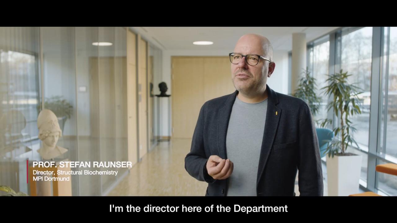
“Electron tomography cannot only be used for our muscle research, but of course, you can imagine for cellular processes like septations or segregation of chromosomes or all kinds of different cellular aspects that really happen inside the cell and that you cannot reconstitute easily.”
Professor Stefan Raunser
Director of the Department of Structural Biochemistry
Max Planck Institute of Molecular Physiology
Dortmund, Germany
“We want to understand structure and function of cellular processes that are relevant for human health across scales. We can do this now here at Human Technopole. We can go from following the activity of macromolecular complexes by light microscopy to investigating their molecular structures within cells by cryo-ET or in isolation at higher resolution by single particle analysis to X-ray crystallography. We can cover the complete range of resolution.”
Professor Gaia Pigino
Human Technopole, Associate head of the Structural Biology Research Centre
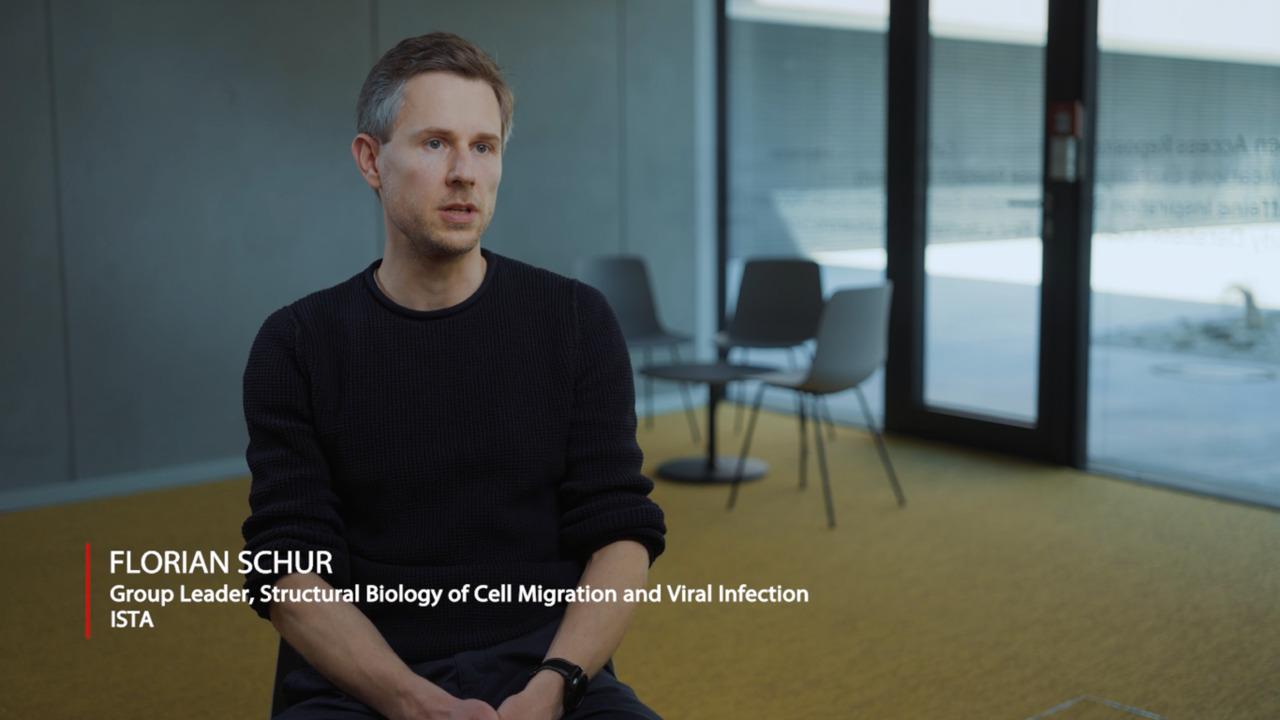
“So with the workflow that we have now fully established at ISTA, we can really address a multitude of questions going from cell biology to high resolution structural biology all within a native context.”
Professor Florian Schur
Institute of Science and Technology Austria
Group Leader
Structural Biology of Cell Migration and viral Infection
ISTA
"One of the things that I find exciting about Arctis [Cryo-PFIB] is the degree to which it is an automated workflow. And so, this ability to have it running essentially unsupervised and produce high quality, large numbers of lamella is transformational."
Jim Naismith
Director of The Rosalind Franklin Institute

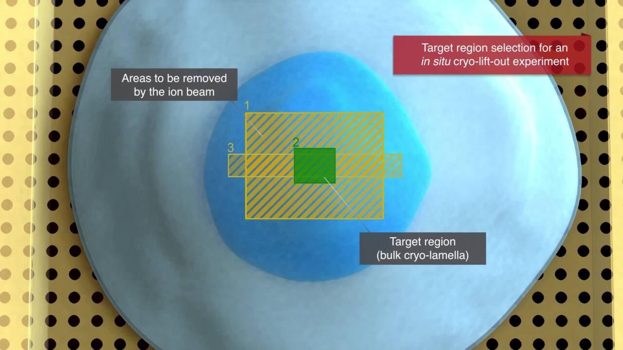
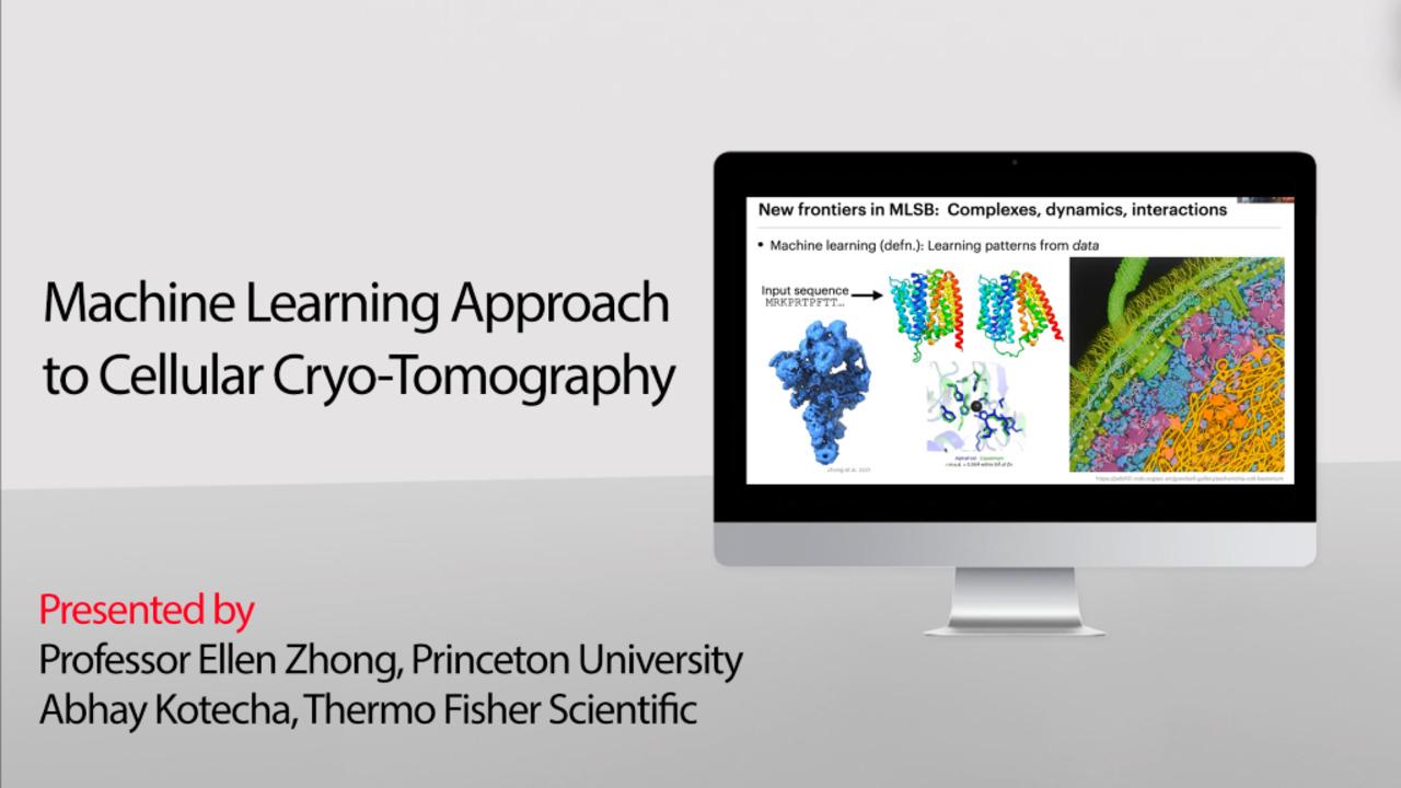
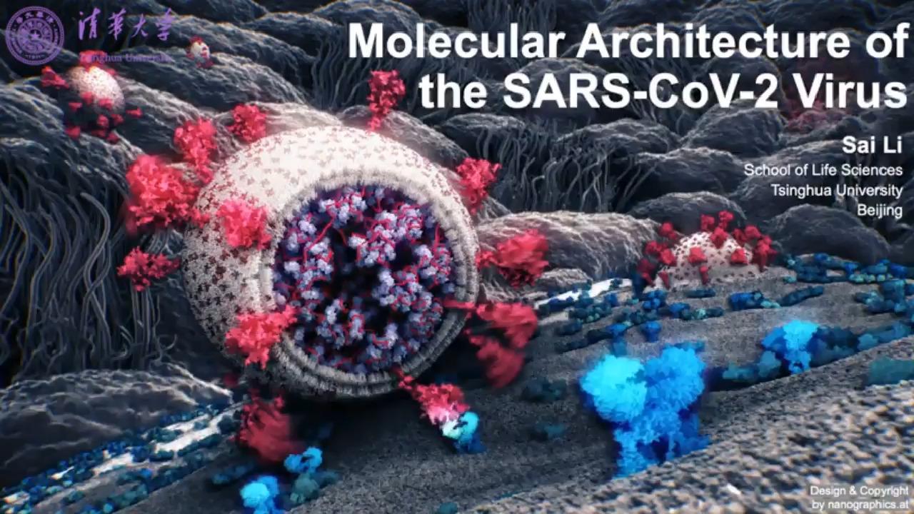
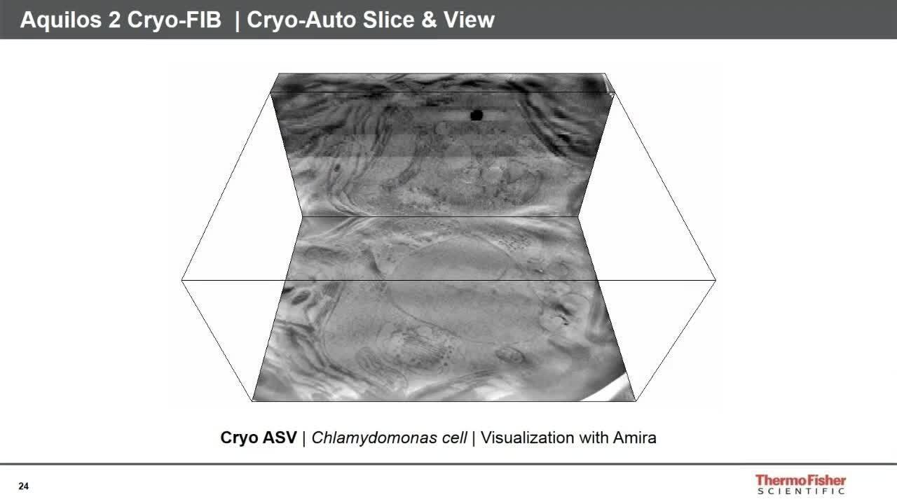
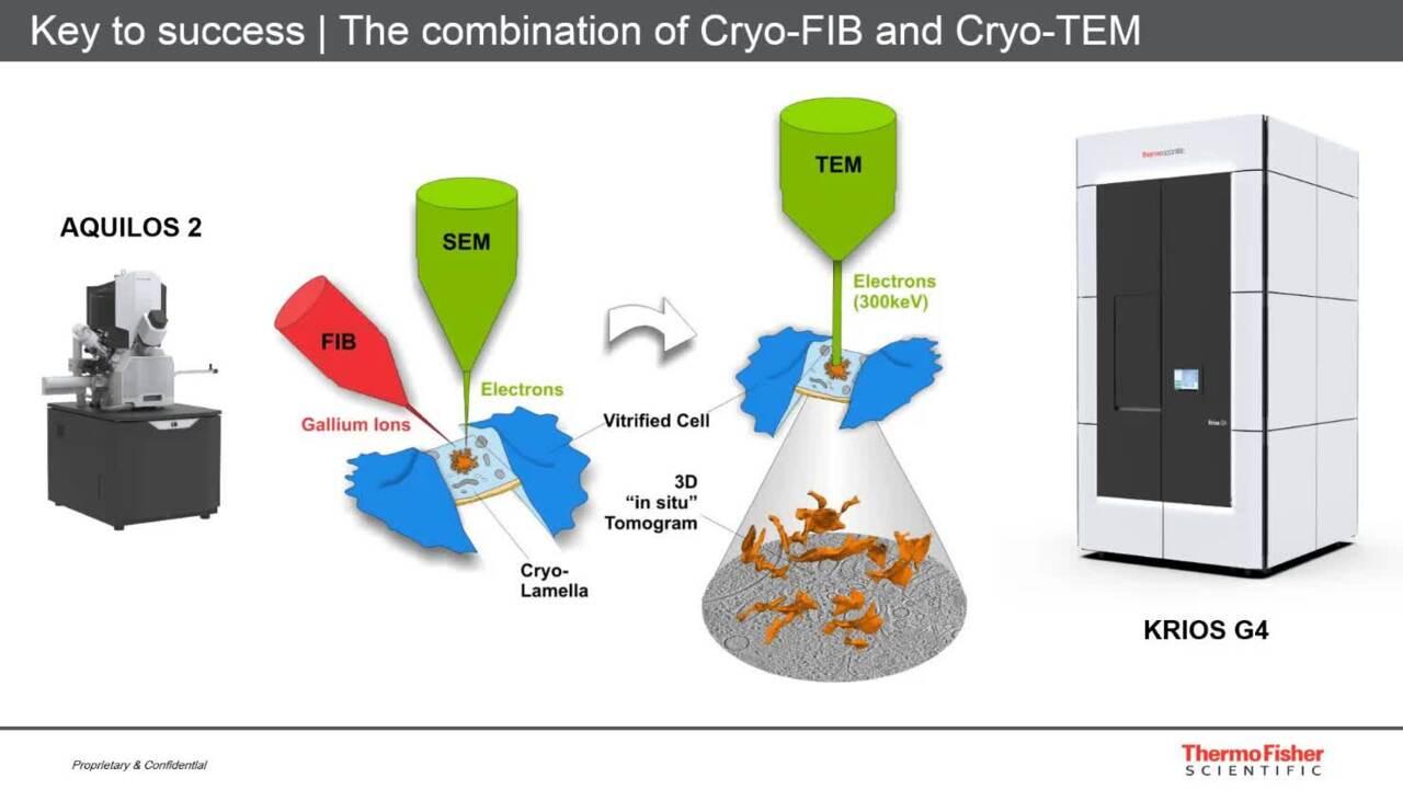
Cryo-electron tomography allows you to visualize macromolecular structures in situ, inside the cell. Vitreous frozen cells are first thinned with a focused ion beam and then imaged in three dimensions using a transmission electron microscope.
In this webinar tutorial, you will find:
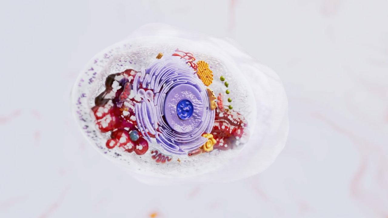
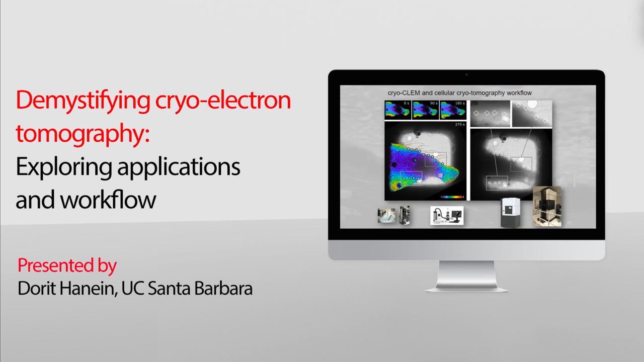
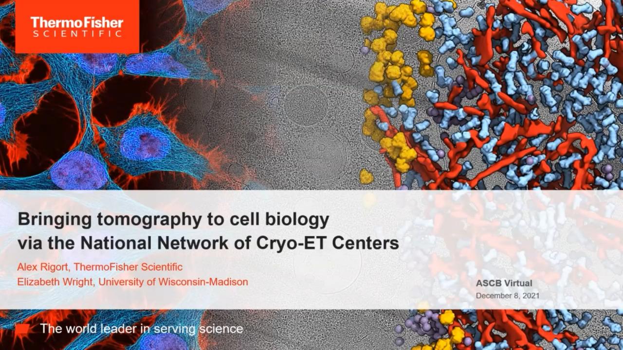
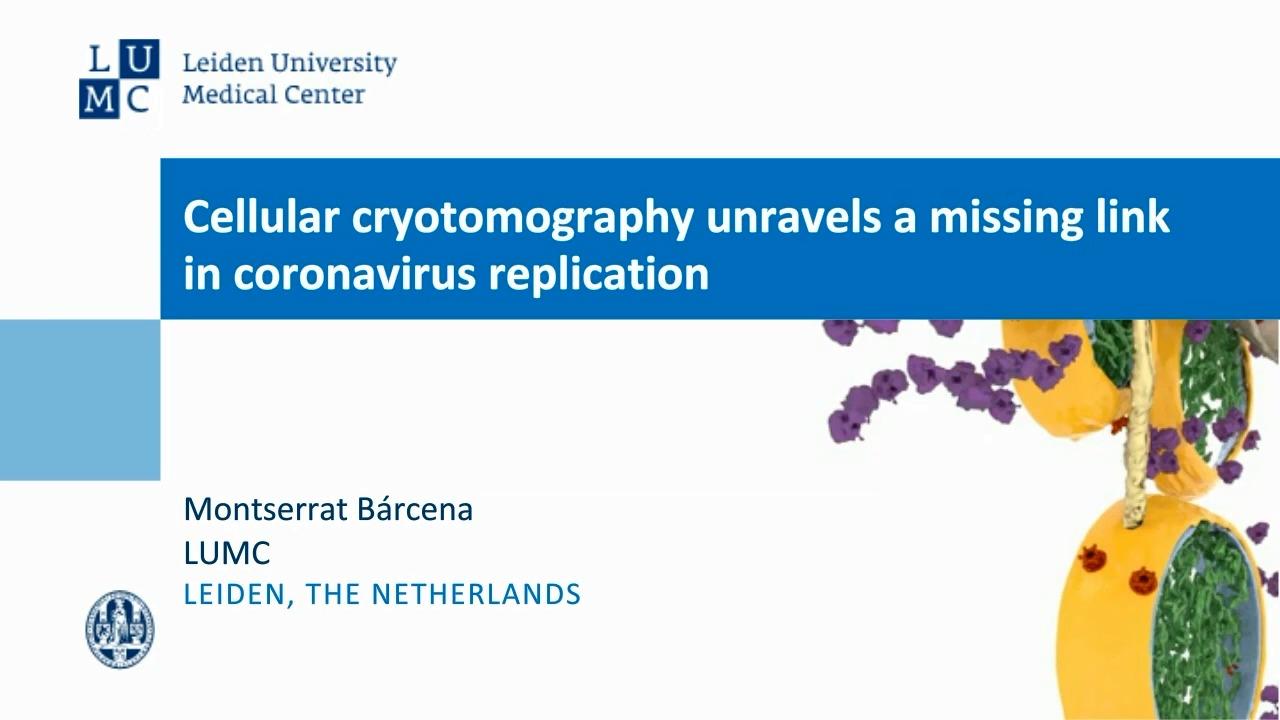
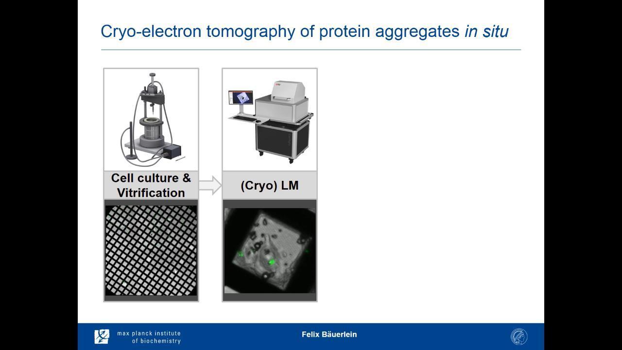
Cellular cryo-electron tomography has changed the way biological systems can be analyzed and modelled. Using this approach, intact cells can now be imaged in their native state and at high resolution, allowing both the quantitative analysis of cellular constituents, as well as modelling of dynamic cellular processes. Some of these might be rare or localized to specific sites within a cell.
Join this high-resolution journey from the membrane to the center of the cell to learn:
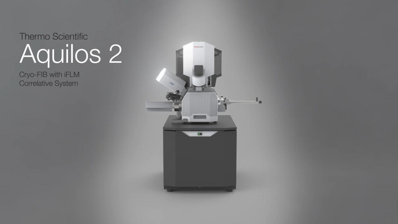
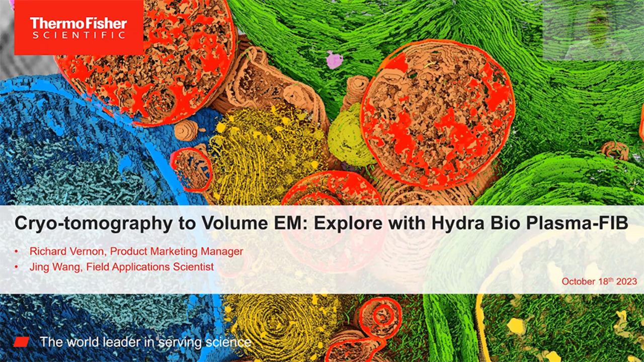
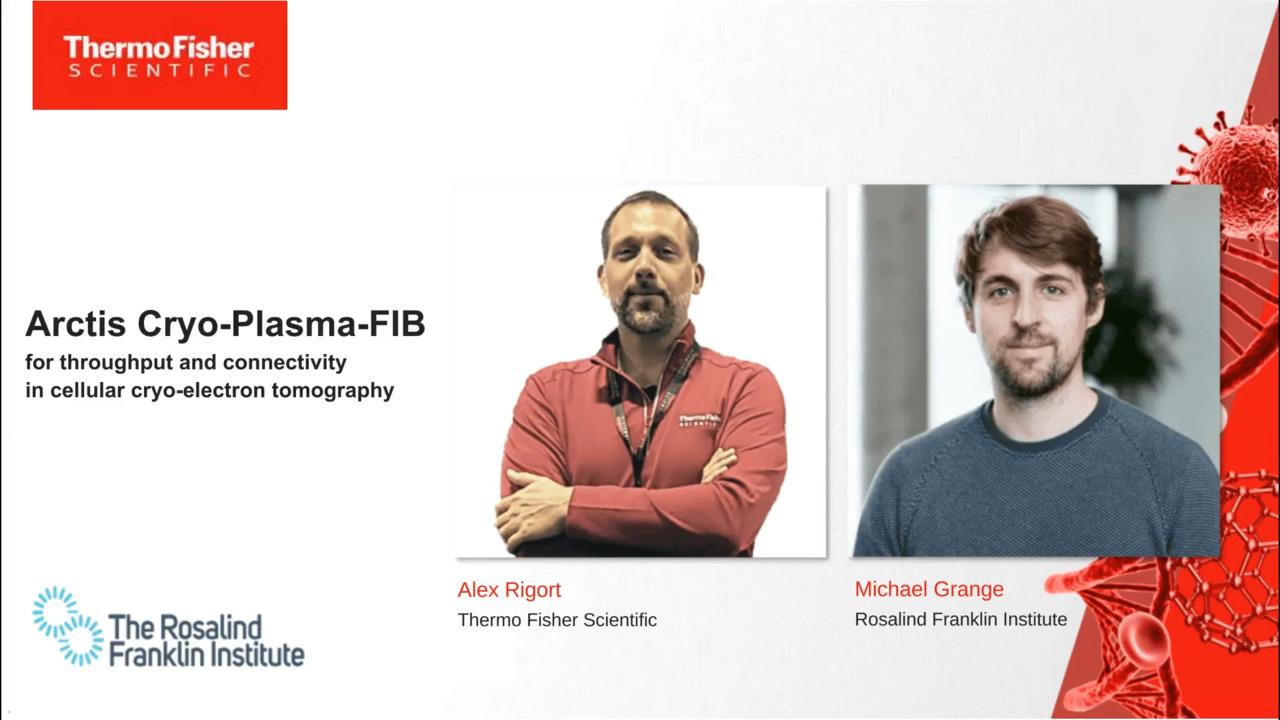
Cellular cryo-electron tomography is a high-resolution technique that enables imaging of a cell’s molecular machinery at close-to-native conditions. With the advent of dedicated cryo-FIB instruments, modern energy filters, fast direct electron detectors, and advanced data acquisition, exciting new opportunities have opened for cryo-electron tomography to shed light on fundamental questions in cell and in situ structural biology. In this webinar, we present our next-generation Thermo Scientific Arctis Cryo-PFIB, which pioneers the systematic production of lamellae from cellular samples for cryo-electron tomography. The new microscope features a state-of-the-art plasma ion source, an automatic sample loading system (Autoloader), integrated fluorescence microscopy for correlative imaging, and direct connectivity to the cryo-transmission electron microscope. This enables the production of cryo-lamellae with higher throughput and better productivity than ever before.
There are nearly 300 world-class cryo-EM facilities where you can advance your research by obtaining cryo-EM structures and proof of concept data using your own samples. Individual facilities also offer different levels of service, which may include sample preparation, grid screening, and cryo-EM training.
For Research Use Only. Not for use in diagnostic procedures.