Search Thermo Fisher Scientific
- Order Status
- Quick Order
-
Don't have an account ? Create Account
Search Thermo Fisher Scientific
Cellular cryo-electron tomography (cryo-ET) is a label-free cryogenic imaging technique that provides 3D datasets of organelles and protein complexes at nanometer resolution in their physiological environments. This is done by opening windows into the cell with focused ion beam (FIB) milling of a cryogenically frozen (vitrified) cell. A series of 2D images is taken of this thinned cellular sample (cryo-lamella) then reconstructed into a 3D dataset. Such high-resolution 3D images of the interior of cells provide new insights into cellular function and sheds light on the arrangement and structure of native protein complexes.
Cryo-electron tomography, a cryo-electron microscopy (cryo-EM) technique, provides 3D snapshots of proteins at work within their functional cellular environments, allowing users to see and understand how they, and other molecules, work together to carry out major processes in a cell. This is because cryo-ET delivers both structural information about individual proteins as well as their spatial arrangements within the cell, making it a truly unique technique. Cryo-ET has enormous potential for cell biology as it bridges the gap between light microscopy and near-atomic resolution techniques like single-particle analysis. Cryo-electron tomography data can be collected with the same transmission electron microscopes as single-particle analysis data.
Cryo-ET provides label-free, fixation-free, 3D nanometer-resolution imaging of cells’ inner workings. Using a correlative light and electron (CLEM) approach allows targeting of tagged proteins by fluorescence microscopy before subsequent higher resolution imaging with cryo-EM. Many cells are too thick for electrons, so the vitrified cells must be thinned with a cryo-focused ion beam microscope (cryo-FIB) prior to imaging in transmission electron microscope.

Cell culture: Cells prepared by routine culture methods are grown on carbon-coated gold electron microscopy (EM) grids.
Vitrification: Cells are either vitrified through plunge-freezing (like single particle specimens) or High Pressure Freezing (HPF). The water in the sample freezes rapidly and does not crystallize, thus avoiding the molecular-scale disruption (by formed ice crystals) that would occur with a normal slow freezing process.
Localization by fluorescence: Using cryo-correlative microscopy, the sample is transferred to a cryo-fluorescence light microscope (cryo-FLM), with which structures of interest are identified. A dedicated cryo-FLM stage keeps the sample in its vitrified state during cryo-fluorescence imaging.
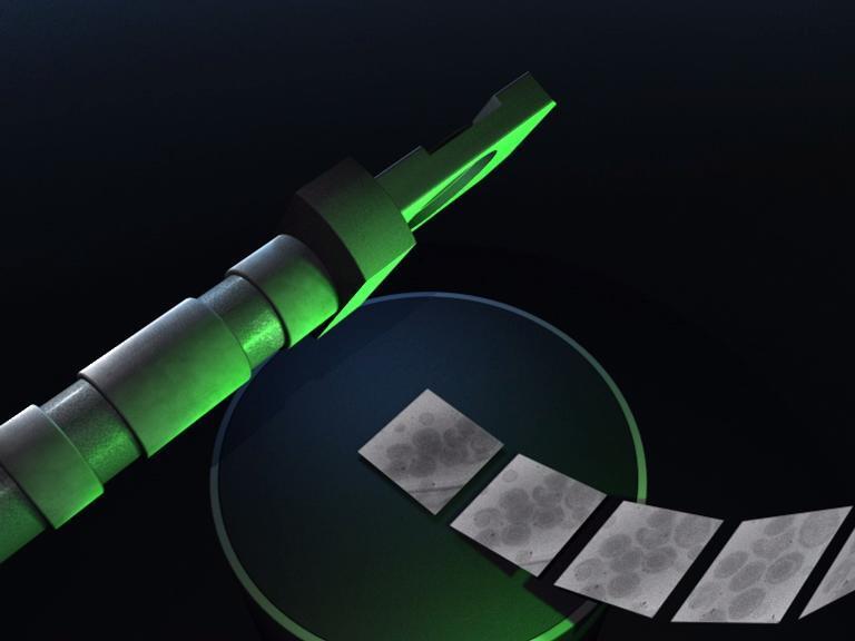
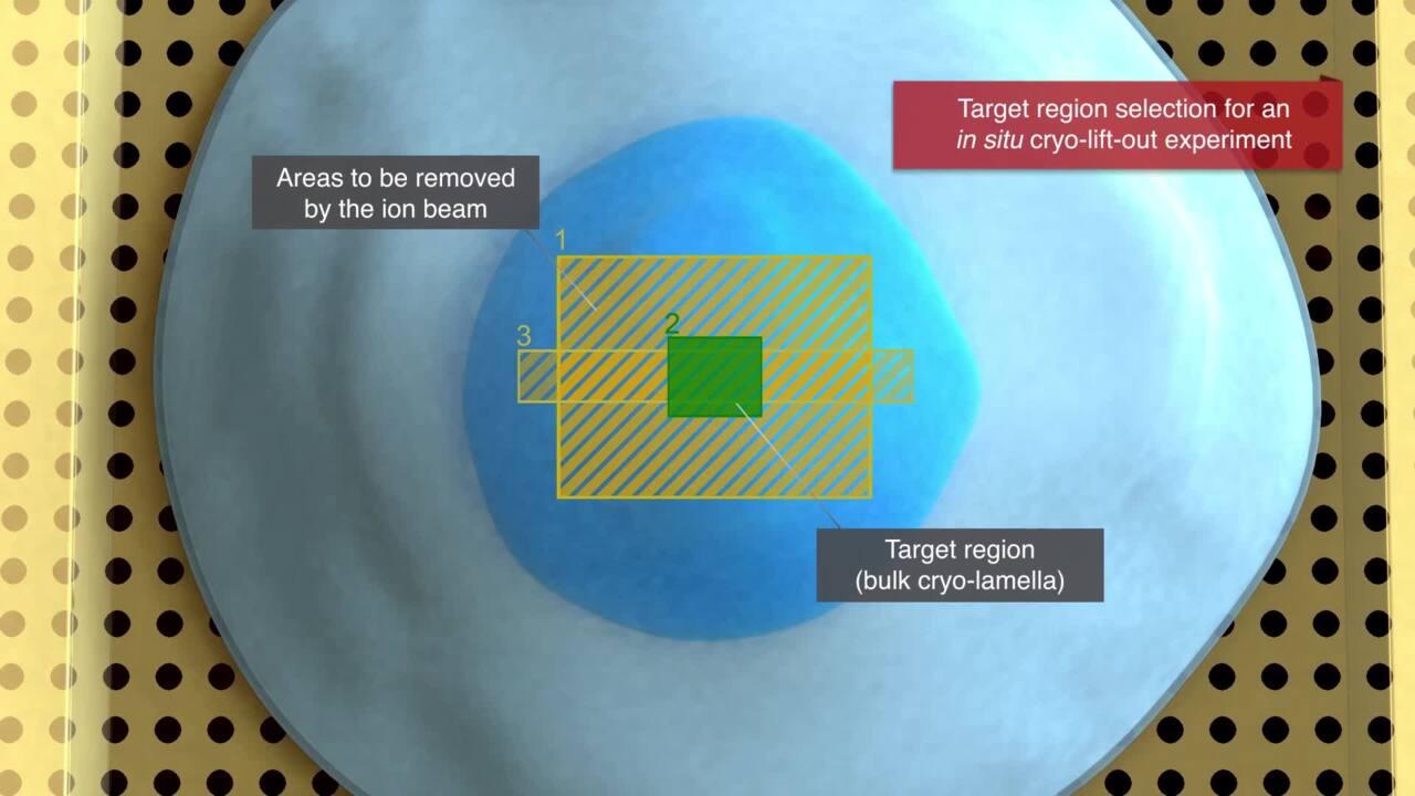
Thinning by milling: A dedicated cryo-FIB prepares a thin, uniform lamella at the vitreous temperature (approximately -170°C).
Imaging by TEM: During cryo-ET, the sample is tilted in known increments about an axis. The individual projection images from the tomographic tilt series are then combined computationally in a procedure known as back-projection, which creates the 3D tomographic volume.
Reconstruction and visualization: The 3D tomogram featuring cellular structures can be segmented and colored in a variety of ways to enhance its display and presentation. From the tomogram, small subsets of data containing the structures of interest can be computationally extracted and subjected to image processing methods.
Cryo-electron tomography has been applied to many different sample types, from single molecules, to protein complexes, viruses, bacteria, and cells, to tissue cells and large tissue samples. Cells are either vitrified through plunge-freezing (as in single particle analysis) or high-pressure freezing (HPF). The water in the sample freezes rapidly and does not crystallize, thus avoiding the molecular-scale disruption (caused by ice crystal formation) that would occur with a normal slow freezing process. Samples are carried on grids, which are designed for the transmission electron microscope. Depending on the size of the sample, plunge freezing is all that is needed. However, for larger samples like cells or tissues, samples must be thinned by cryo-FIB milling so that electrons can pass through the sample.

Cryo-ET is used to obtain 3D information from vitrified samples. Depending on the size of the sample, a thinning step is included in the sample preparation using focused ion beam (FIB) or plasma ion beam milling to produce cryo-lamella that can be penetrated by electron beams.
Light microscopy, particularly fluorescence microscopy, is a cornerstone of modern cell biology. Advances in optics, as well as computational tools, have made it possible to view systems in a range of relevant scales. It allows for the identification of proteins and molecules based on bound fluorescent tags. Structures that are not fluorescently labeled, however, cannot be visualized, meaning the context surrounding the fluorescent label is lost. Moreover, the presence of certain chemical agents needed for some fluorescence modalities to work can also affect the native structure. Locating the structure of interest can be difficult in the vast complexity of the natural cellular environment. However, using cryo-correlative microscopy (cryo-CLEM), the structures of interest are identified in the cryo-fluorescence light microscope. The iFLM (Integrated Fluorescence Light Microscope) Correlative System delivers the localization of fluorescent targets inside the chamber, allowing users to check the fluorescence signal inside milled lamellae, eliminating extra sample transfer steps and correlate two imaging modalities directly within one system. The iFLM Correlative System is available on the Thermo Scientific Aquilos 2 Cryo-FIB, Thermo Scientific Helios Hydra Cryo-Plasma-FIB, and Thermo Scientific Arctis Cryo-Plasma-FIB.
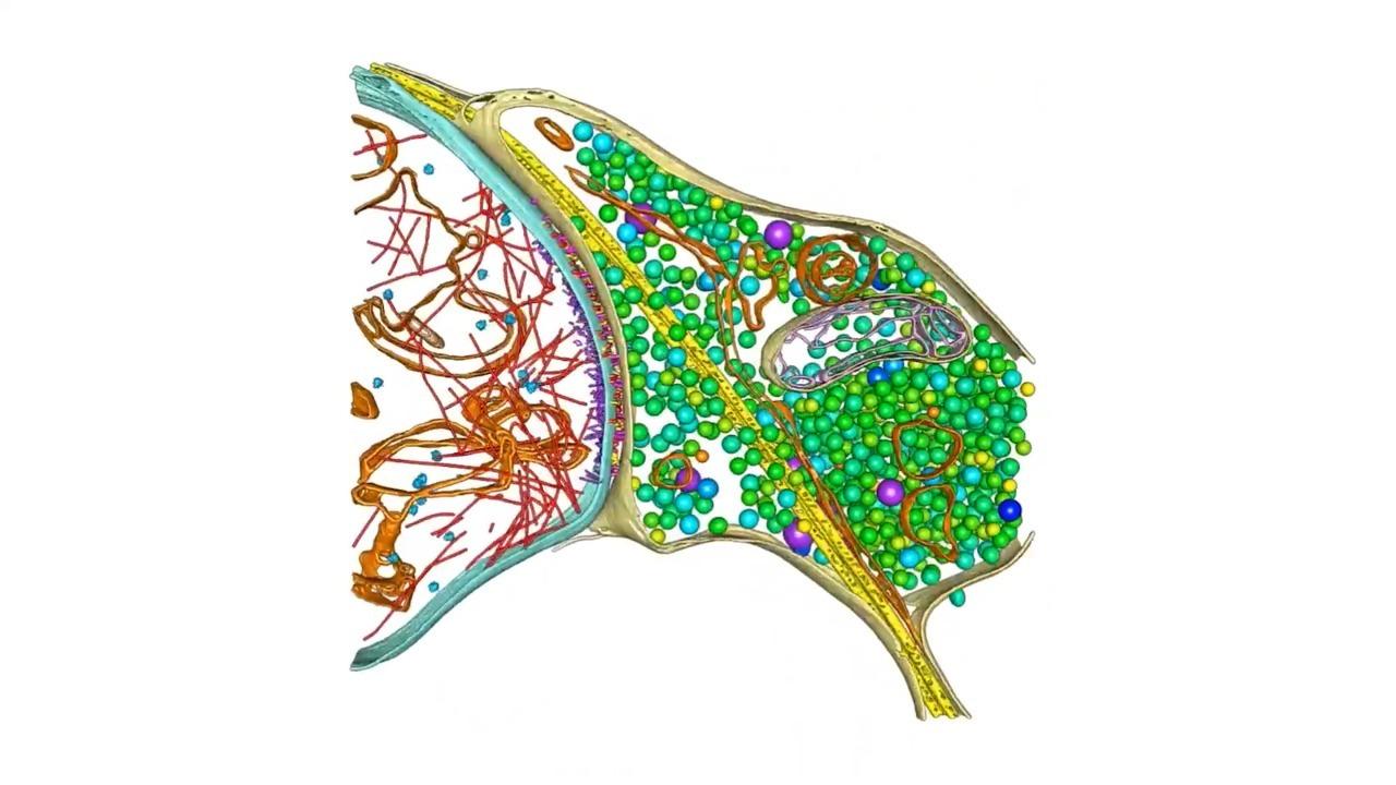
 Tomography structure of a HeLa cell. Cryo-electron tomography provides a close up containing vital contextual information.


Cryo-lamella prepared with the Arctis Cryo-Plasma-FIB (right). Overlay of SEM image with fluorescence image obtained with the iFLM Correlative System (left)
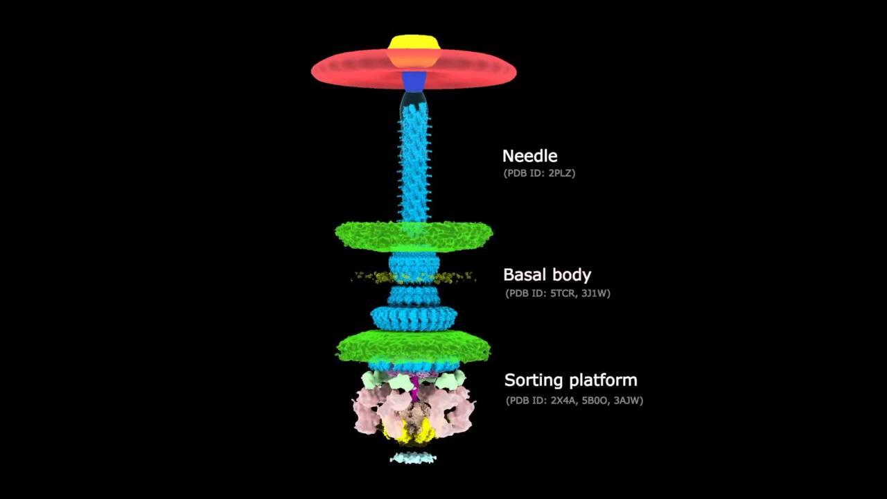
Cryo-ET is the only method that can achieve near-atomic resolutions while observing cellular structures in their native context. The resolution range for cryo-ET is large, bridging the gap between micron-level resolution, where cellular structure can be visualized, and nanometer resolution, where protein complexes can be visualized.

Reliable results from cryo-ET rely on high-quality cryo-lamella preparation. When used with light microscopy, which provides the location of a labeled protein of interest, cryo-ET brings together the best of both worlds: localization, specificity, and nanometer resolution. A new integrated cryo-ET workflow from Thermo Fisher Scientific illustrates how these two techniques can be combined to achieve results.


For Research Use Only. Not for use in diagnostic procedures.