Search Thermo Fisher Scientific
Verios 5 XHR Scanning Electron Microscope
The Verios 5 XHR SEM offers subnanometer resolution over the full 1 keV to 30 keV energy range with excellent materials contrast. Unprecedented levels of automation and ease-of-use make this performance accessible to users of any experience level.
Scanning electron microscopy characterization
- High resolution nanomaterial imaging with the UC+ monochromated electron source for sub-nanometer performance from 1-30 kV.
- High contrast on sensitive materials with excellent performance down to 20 eV landing energy and high-sensitivity in-column and below-the-lens detectors and signal filtering for low-dose operation and optimal contrast selection.
- Greatly reduced time to nanoscale information for users with any experience level using the Elstar electron column featuring SmartAlign and FLASH technologies.
- Consistent measurement results with ConstantPower lenses, electrostatic scanning and a choice of two piezoelectric stages.
- Flexibility for accessories with a large chamber.
- Unattended SEM operation with Thermo Scientific AutoScript 4 Software, an optional Python-based application programming interface.
SmartAlign technology
SmartAlign technology eliminates the need for any user alignments of the electron column, which not only minimizes maintenance, but also increases your productivity.
Innovative electron optics
Including Thermo Scientific’s patented UC+ gun (monochromator), ConstantPower lenses and electrostatic scanning for accurate and stable imaging.
Sub-nanometer resolution
Elstar Schottky monochromated (UC+) FESEM technology and performance with sub-nanometer resolution from 1 to 30 keV.
Consistent measurement results
The Verios is ideally suited to lab-based metrology applications, with the ability to calibrate to a NIST certified standard at high magnification.
Low dose operation and optimal contrast selection
Advanced suite of high-sensitivity, in-column & below-the-lens detectors and signal filtering for low dose operation and optimal contrast selection.
Easy access to beam landing energies
As low as 20 eV with very high resolution for true surface characterization.
Unattended SEM operation
With AutoScript 4 Software, an optional Python-based application programming interface (API).
Large chamber
With a choice of two precise and stable piezo-driven stages.
Electron beam resolution |
| |
Standard detectors | ETD, TLD, MD, ICD, beam current measurement, Nav-Cam+, IR-camera | |
Optional detectors | Optional detectors | EDS, EBSD, RGB cathodoluminescence, Raman, WDS, and more | |
Stage bias (beam deceleration, optional) | Up to -4000 V, included as standard | |
Sample cleaning | Integrated plasma cleaner, included as standard | |
Sample manipulation | Verios 5 UC
| Verios 5 HP
|
Chamber | 379 mm inside width, 21 ports | |
Software options |
| |
Webinar introducing the new Verios 5 XHR SEM
This webinar will present Thermo Scientific technology advances in electron source, electron column, detector and the user interface that enables routine ultra-low voltage SEM imaging and characterization. By watching the webinar, you’ll learn how to:
- Find out which level of information high-performance SEM is able to provide for nanomaterial samples
- Understand how different technologies on the Verios 5 work in unison for exceptional low-V performance
- Discover how user interface automation brings expert results to all users
Webinar: Scanning electron microscopy: selecting the right technology for your needs
This on-demand webinar has been designed to help you decide which SEM best meets your unique needs. We present an overview of Thermo Fisher Scientific SEM technology for multi-user research labs and focus on how these wide-ranging solutions deliver performance, versatility, in situ dynamics and faster time to results. Watch this webinar if you are interested in:
- How the needs for different microanalysis modalities are met (EDX, EBSD, WDS, CL, etc.).
- How samples are characterized in their natural state without the need for sample preparation.
- How new advanced automation allows researchers to save time and increase productivity.

Fundamental Materials Research
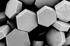
Novel materials are investigated at increasingly smaller scales for maximum control of their physical and chemical properties. Electron microscopy provides researchers with key insight into a wide variety of material characteristics at the micro- to nano-scale.

Semiconductor Pathfinding and Research
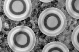
Advanced electron microscopy, focused ion beam, and associated analytical techniques for identifying viable solutions and design methods for the fabrication of high-performance semiconductor devices.

Yield Ramp and Metrology
We offer advanced analytical capabilities for defect analysis, metrology, and process control, designed to help increase productivity and improve yield across a range of semiconductor applications and devices.

Semiconductor Failure Analysis
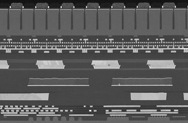
Increasingly complex semiconductor device structures result in more places for failure-inducing defects to hide. Our next-generation workflows help you localize and characterize subtle electrical issues that affect yield, performance, and reliability.

Physical and Chemical Characterization
Ongoing consumer demand drives the creation of smaller, faster, and cheaper electronic devices. Their production relies on high-productivity instruments and workflows that image, analyze, and characterize a broad range of semiconductor and display devices.

Energy Dispersive Spectroscopy
Energy dispersive spectroscopy (EDS) collects detailed elemental information along with electron microscopy images, providing critical compositional context for EM observations. With EDS, chemical composition can be determined from quick, holistic surface scans down to individual atoms.

Imaging Hot Samples
Studying materials in real-world conditions often involves working at high temperatures. The behavior of materials as they recrystallize, melt, deform, or react in the presence of heat can be studied in situ with scanning electron microscopy or DualBeam tools.

Multi-scale analysis
Novel materials must be analyzed at ever higher resolution while retaining the larger context of the sample. Multi-scale analysis allows for the correlation of various imaging tools and modalities such as X-ray microCT, DualBeam, Laser PFIB, SEM and TEM.

Cathodoluminescence
Cathodoluminescence (CL) describes the emission of light from a material when it is excited by an electron beam. This signal, captured by a specialized CL detector, carries information on the sample’s composition, crystal defects, or photonic properties.
SEM Metrology
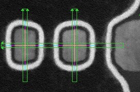
Scanning electron microscopy provides accurate and reliable metrology data at nanometer scales. Automated ultra-high-resolution SEM metrology enables faster time-to-yield and time-to-market for memory, logic, and data storage applications.

Energy Dispersive Spectroscopy
Energy dispersive spectroscopy (EDS) collects detailed elemental information along with electron microscopy images, providing critical compositional context for EM observations. With EDS, chemical composition can be determined from quick, holistic surface scans down to individual atoms.

Imaging Hot Samples
Studying materials in real-world conditions often involves working at high temperatures. The behavior of materials as they recrystallize, melt, deform, or react in the presence of heat can be studied in situ with scanning electron microscopy or DualBeam tools.

Multi-scale analysis
Novel materials must be analyzed at ever higher resolution while retaining the larger context of the sample. Multi-scale analysis allows for the correlation of various imaging tools and modalities such as X-ray microCT, DualBeam, Laser PFIB, SEM and TEM.

Cathodoluminescence
Cathodoluminescence (CL) describes the emission of light from a material when it is excited by an electron beam. This signal, captured by a specialized CL detector, carries information on the sample’s composition, crystal defects, or photonic properties.
SEM Metrology
Scanning electron microscopy provides accurate and reliable metrology data at nanometer scales. Automated ultra-high-resolution SEM metrology enables faster time-to-yield and time-to-market for memory, logic, and data storage applications.


