Electron Microscopy for the Life Sciences

Going beyond the beam to empower life science discoveries

From enabling scientific discoveries to breakthrough drug discovery, our electron microscopy solutions are designed to reveal molecular details at biologically relevant resolutions, helping you understand cellular ultrastructure, protein function, disease mechanisms, and facilitate effective drug design. We deliver the products, services, expertise, and technical support you need to push the boundaries of what’s possible in your research and your pursuit to see the unseen. 

Cryo-Electron Microscopy | Cryo EM

Cryo FIB | Focused Ion Beam SEM

走査電子顕微鏡

TEMイメージング

Electron microscopy innovations for the life sciences

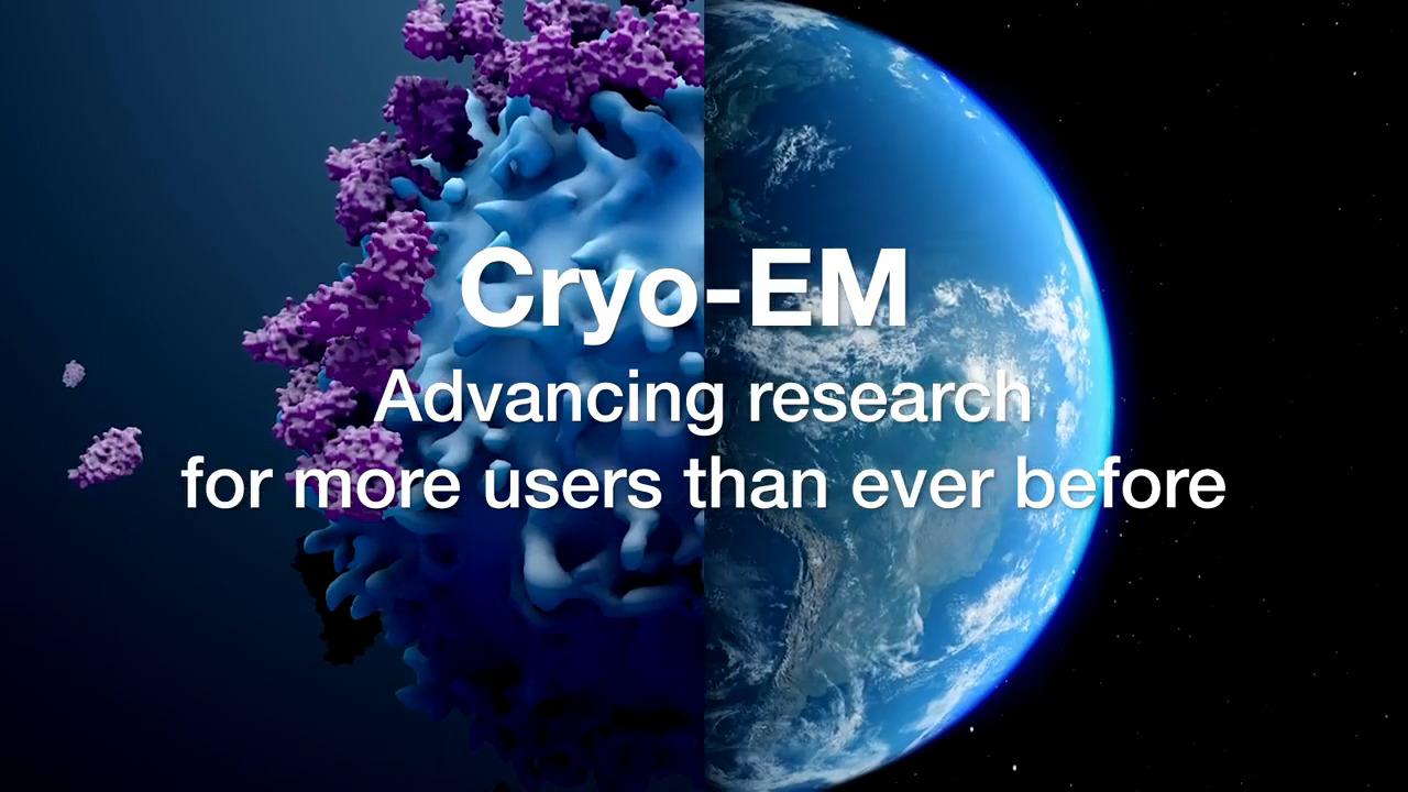
Cryo-electron microscopy (cryo-EM) is rapidly becoming the method of choice for biochemistry labs around the world, helping accelerate research. With cryo-EM, molecular details can be seen at biologically relevant resolutions, providing insights into protein function and disease mechanisms, and facilitating effective drug design. Recent advances in instrument design and automation mean cryo-EM is easier to use and more cost effective than ever before. With cryo-EM, you can gain deeper insights into complex biological processes and pathways. While researchers use a variety of techniques to determine protein structure, cryo-EM can provide details on protein function that other methods simply cannot.


Life science electron microscopy techniques and workflows

Single particle analysis

Single particle analysis is a cryo-electron microscopy technique that enables structural characterization at near-atomic resolutions, unraveling dynamic biological processes and the structure of biomolecular complexes/assemblies.

Cryo-electron tomography

Cryo-electron tomography delivers both structural information about individual proteins as well as their spatial arrangements within the cell, bridging the gap between light microscopy and near-atomic-resolution techniques like single particle analysis.

Rational drug design

Discover how cryo-EM is being used for structure-based drug design to unlock new targets and modalities.

Microcrystal electron diffraction (MicroED)

MicroED is an exciting technique with applications in the structural determination of small molecules and protein. With this method, atomic details can be extracted from individual nanocrystals (<200 nm in size), even in a heterogeneous mixture.

Volume electron microscopy

Volume electron microscopy (vEM) is a group of techniques that reveals the 3D ultrastructure of cells and tissues at micron to millimeter volume scales, at nanometer-level resolutions, and even at native state under cryogenic conditions.

Cryo-electron microscopy sample preparation

Successful protein preparation lies at the base of any successful structural biology technique. For cryo-electron microscopy, a wide range of solutions is required to achieve the highest sample quality before freezing of the specimen on the EM grid.

Visualization and analysis software

A powerful, flexible, versatile 2D-5D software platform for visualization, processing, and analysis of microscopy imaging.

Integrative structural biology

To understand protein function, you need complex and structure information beyond individual proteins. Integrative structural biology combines mass spectrometry and cryo-EM for the determination of large dynamic complex structures.

Getting started in Cryo-EM

EM Resource Center