Search Thermo Fisher Scientific
Quattro Environmental Scanning Electron Microscope
The Thermo Scientific Quattro ESEM combines all-around performance in imaging and analytics with a unique environmental mode (ESEM) that allows samples to be studied in their natural state. It is ideal for a wide variety of academic, industrial, and government labs that want the versatility and ease of use needed for multiple users of different experience levels and disciplines on a platform that also supports unique in situ experiments. The Quattro ESEM features a field emission gun (FEG), which ensures excellent resolution. Meanwhile, its three vacuum modes (high vacuum, low vacuum, and ESEM) provide the flexibility to accommodate the widest range of samples of any SEM available, including those that are outgassing or otherwise not vacuum-compatible.

Learn more about this product in our on-demand webinar
Discover how to analyze liquids and hydrated samples in 3D using environmental scanning electron microscopy.
Best in class for in situ imaging capabilities, dynamic experiments with excellent analytical capabilities
The Quattro ESEM is a highly flexible platform for in situ dynamic experiments. The Quattro ESEM's environmental SEM capability allows scientists to study materials in a range of conditions, such as wet/humid, hot, or reactive environments, as they develop new materials and products. It supports cooling and heating experiments both in high vacuum and low vacuum to accommodate the widest range of needs. Cooling experiments on wet materials are possible with the Peltier cooling stage or if the transmission mode is needed, with Thermo Scientific WetSTEM Technology. Additionally, the Quattro ESEM provides several different possibilities to heat the samples of interest, both bulk and powders. Users can perform dynamic experiments at high temperatures both in high vacuum (with the high-vacuum heating stage), in low-vacuum/ESEM mode (with the two ESEM heating stages for temperatures up to 1,000°C and 1,400°C), and on a localized area for better control of the temperature change (with the Thermo Scientific µHeater Holder).
Thanks to the Quattro ESEM chamber size, it accommodates a wide range of accessories. Analytical capabilities include energy dispersive X-ray spectroscopy (EDS) with ports for 180-degree dual EDS attachment, electron backscatter diffraction (EBSD) coplanar with EDS, and wavelength dispersive X-ray spectroscopy (WDS). Furthermore, the Quattro ESEM comes with the new Thermo Scientific ChemiSEM Technology, a unique live elemental imaging capability that is fully integrated into the SEM user interface, providing always-available compositional information through the most intuitive interface.
Diverse material application observed with the Quattro ESEM
Dynamic in situ experiments
In situ study of materials in their natural state: unique high-resolution FEG-SEM with environmental mode (ESEM). In situ analysis at temperatures ranging from -165°C to 1,400°C with a range of cryo, Peltier, and heating stages.
Peltier cooling experiment of sodium sulfate crystals in sandstone.
In situ cooling experiment conducted on a sandstone sample with crystals of sodium sulfate. The target of the experiment was to study the sodium sulfate behavior when completely hydrated. The pressure was varied up to 700 Pa, maintaining the temperature at 2˚C. Sample courtesy of Institute of Theoretical and Applied Mechanics of the Academy of Science, Czech Republic.
μHeater Holder heating experiment of magnetite and hematite.
Heating experiment run with the μHeater Holder. The video shows the heating of a mix of magnetite and hematite from 40˚C up to 1,000˚C.
Wide range of information
Observe all information from all samples with simultaneous SE and BSE imaging in every mode of operation. Several additional detectors, such as the STEM3+ detector and the retractable RGB cathodoluminescence (CL) detector are available to accommodate every user’s need and provide a complete set of information from a wide range of materials.
Excellent analytical capabilities
Excellent analytical capabilities with a flexible chamber that allows multiple EDS, EBSD, or WDS detectors. Elemental information at your fingertips with ChemiSEM Technology, which provides live, quantitative, elemental mapping for unprecedented time to result and ease of use. Excellent analysis of non-conductive samples: accurate EDS and EBSD are enabled in low vacuum with the Quattro ESEM's through-the-lens pumping.
Minimize sample preparation time with the unique combination of high-vacuum, low-vacuum, and environmental modes
Ease of use with innovative options and advanced automation
Easy to use, intuitive software with User Guidance. The unique Undo function permits efficient exploration of imaging conditions and allows you to work faster with fewer mouse clicks.
Advanced automation
Advanced automation is provided in different ways, with either Thermo Scientific AutoScript 4 Software or Thermo Scientific Maps Software, depending on the specific application needs.
AutoScript 4 Software offers control of the Quattro ESEM. Take advantage of a Python-based application programming interface (API) to optimize your in situ cooling or heating experiments or record and monitor dynamic parameters such as temperature, stage position, or pressure.
Maps Software automates large-area acquisition on multiple samples with up to four different simultaneous signals. In addition to a clear increase in the system productivity, it offers a multi-scale, multi-layered visualization environment in which 2D and 3D data and imagery can be imported from any source to correlate different modalities.
ESEM heating stage experiment on a silver wire, heated from 300˚C to 530˚C. AutoScript 4 Software was used to obtain a live drift compensation through a combination of beam shift and stage moves. The result was a stable image of the silver wire for the whole duration of the movie, even when the wire changed location over the substrate.
| Resolution |
|
| Standard detectors |
|
| Optional detectors |
|
| ChemiSEM Technology (optional) |
|
| Stage bias (beam deceleration, optional) |
|
| Low vacuum mode |
|
| Stage |
|
| Standard sample holder |
|
| Chamber |
|
| In situ accessories (optional) |
|
| Software options |
|
ESEM cooling experiment on coated filter paper, showing the paper’s behavior when fully hydrated.
WetSTEM cooling experiment on a flower and pollen sample. Thanks to the flexible design of this stage, it allows the characterization both in transmission mode and in top-down mode.
Peltier cooling experiment on food gelatin. The video shows the microstructural changes of the gelatine when frozen down to -10˚C.
Heating experiment ran with the μHeater system. The video shows the heating of a mix of magnetite and hematite from 40˚C up to 1000˚C.
Metals heating with the high vacuum heating stage. the video shows how the melted gold moves over the other materials while the heating progresses.
Cooling experiment conducted with the Peltier cooling stage. It shows the hydration of mold spores from a cheese sample.
In-situ cooling experiment conducted on a sandstone sample with crystals of sodium sulphate, to study its behavior when completely hydrated. Sample courtesy of Institute of Theoretical and Applied Mechanics of the Academy of Science, Czech Republic.
In-situ cooling experiment conducted on a sandstone sample with crystals of sodium sulphate, to study its behavior when completely hydrated. Sample courtesy of Institute of Theoretical and Applied Mechanics of the Academy of Science, Czech Republic.
ESEM heating stage experiment on a silver wire, heated from 300˚C to 530˚C. Autoscript 4 was used to obtain a live drift compensation through a combination of beam shift and stage moves.
Heating of a solder wire from 200˚C to 350˚C ran with the high vacuum heating stage.
TiAl TNM-B1 alloy heating with the high vacuum heating stage. The alloy was heated from room temperature up to 1100˚C showing oxidation and phase changes on the surface.
μHeater heating experiment on zinc oxide platelets. The progressive heating up to 1000˚C shows the textural changes of the platelet.
Webinar: Scanning electron microscopy: selecting the right technology for your needs
This on-demand webinar has been designed to help you decide which SEM best meets your unique needs. We present an overview of Thermo Fisher Scientific SEM technology for multi-user research labs and focus on how these wide-ranging solutions deliver performance, versatility, in situ dynamics and faster time to results. Watch this webinar if you are interested in:
- How the needs for different microanalysis modalities are met (EDX, EBSD, WDS, CL, etc.).
- How samples are characterized in their natural state without the need for sample preparation.
- How new advanced automation allows researchers to save time and increase productivity.
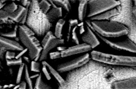
Controle de processo
A indústria moderna exige alta produtividade com qualidade superior, um equilíbrio mantido por meio de um controle de processo robusto. As ferramentas SEM e TEM com software de automação dedicado proporcionam informações rápidas e em várias escalas para monitoramento e aprimoramento de processos.
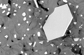
Controle de qualidade
O controle de qualidade e a garantia de qualidade são essenciais na indústria moderna. Oferecemos uma gama de ferramentas de microscopia eletrônica e espectroscopia para análises multidimensionais e multimodais de defeitos, permitindo que você tome decisões confiáveis e informadas para controle e melhoria de processos.
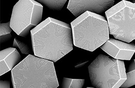
Pesquisa de materiais fundamentais
Novos materiais são investigados em escalas cada vez menores para o máximo controle de suas propriedades físicas e químicas. A microscopia eletrônica fornece aos pesquisadores percepções importantes sobre uma ampla variedade de características materiais em escala micro a nano.
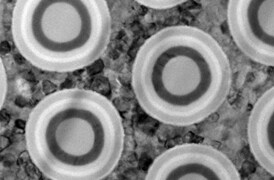
Desenvolvimento e localização de semicondutores
Microscopia eletrônica avançada, feixe de íon focalizado e técnicas analíticas associadas para identificar soluções viáveis e métodos de desenho para a fabricação de dispositivos semicondutores de alto desempenho.
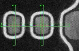
Rampa de escoamento e metrologia
Oferecemos recursos analíticos avançados para análise de defeitos, metrologia e controle de processos projetados para ajudar a aumentar a produtividade e melhorar o rendimento em uma variedade de aplicações e dispositivos semicondutores.
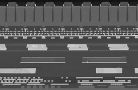
Análise de falha de semicondutores
Estruturas de dispositivos semicondutores cada vez mais complexas resultam em mais locais onde defeitos que induzem falhas podem se ocultar. Nossos fluxos de trabalho de última geração o ajudam a localizar e caracterizar problemas elétricos sutis que afetam o rendimento, o desempenho e a confiabilidade.
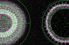
Caracterização física e química
A demanda contínua dos consumidores impulsiona a criação de dispositivos eletrônicos menores, mais rápidos e mais baratos. Sua produção depende de instrumentos e fluxos de trabalho de alta produtividade que fazem imagens, analisam e caracterizam uma ampla gama de semicondutores e dispositivos de exibição.

Espectroscopia por energia dispersiva
A espectroscopia por energia dispersiva (EDS) coleta informações elementares detalhadas juntamente com imagens de microscopia eletrônica, fornecendo contexto de composição crítico para observações EM. Com a EDS, a composição química pode ser determinada a partir de varreduras de superfície rápidas e holísticas que chegam até a átomos individuais.

Geração de imagens de amostras quentes
O estudo de materiais em condições reais muitas vezes envolve trabalhar em altas temperaturas. O comportamento dos materiais à medida que eles se recristalizam, derretem, deformam ou reagem na presença de calor pode ser estudado in situ com microscopia eletrônica de varredura ou ferramentas DualBeam.
_Technique_800x375_144DPI.jpg)
SEM ambiental (ESEM)
A SEM ambiental permite a geração de imagens dos materiais em seu estado nativo. Isso é perfeitamente adequado para pesquisadores acadêmicos e industriais que precisam testar e analisar amostras molhadas, sujas, reativas, gasosas ou não compatíveis com vácuo.

Experimentação in situ
A observação direta e em tempo real de alterações microestruturais com a microscopia eletrônica é necessária para entender os princípios subjacentes de processos dinâmicos, como recristalização, crescimento de grãos e transformação de fases durante o aquecimento, o resfriamento e a umidificação.

Análise de partículas
A análise de partículas tem uma função vital na pesquisa de nanomateriais e no controle de qualidade. A resolução nanométrica e a formação de imagens excelentes da microscopia eletrônica podem ser combinadas com software especializado proporcionando uma rápida caracterização de pós e partículas.

Catodoluminescência
A catodoluminescência (CL) descreve a emissão de luz de um material quando ele é excitado por um feixe de elétrons. Este sinal, captado por um detector CL especializado, carrega informações sobre a composição da amostra, defeitos de cristal ou propriedades fotônicas.

ColorSEM
Usando a EDS (espectroscopia de raios X por energia dispersiva) com quantificação ao vivo, a tecnologia ColorSEM transforma as imagens SEM em uma técnica colorida. Agora, qualquer usuário pode adquirir dados elementares continuamente para obter informações mais completas do que nunca.
Análise e geração de imagens de semicondutores
A Thermo Fisher Scientific oferece microscópios eletrônicos de varredura para todas as funções de um laboratório de semicondutores, desde tarefas gerais de aquisição de imagens até técnicas avançadas de análise de falhas que exigem medições precisas de contraste de tensão.

Espectroscopia por energia dispersiva
A espectroscopia por energia dispersiva (EDS) coleta informações elementares detalhadas juntamente com imagens de microscopia eletrônica, fornecendo contexto de composição crítico para observações EM. Com a EDS, a composição química pode ser determinada a partir de varreduras de superfície rápidas e holísticas que chegam até a átomos individuais.

Geração de imagens de amostras quentes
O estudo de materiais em condições reais muitas vezes envolve trabalhar em altas temperaturas. O comportamento dos materiais à medida que eles se recristalizam, derretem, deformam ou reagem na presença de calor pode ser estudado in situ com microscopia eletrônica de varredura ou ferramentas DualBeam.
_Technique_800x375_144DPI.jpg)
SEM ambiental (ESEM)
A SEM ambiental permite a geração de imagens dos materiais em seu estado nativo. Isso é perfeitamente adequado para pesquisadores acadêmicos e industriais que precisam testar e analisar amostras molhadas, sujas, reativas, gasosas ou não compatíveis com vácuo.

Experimentação in situ
A observação direta e em tempo real de alterações microestruturais com a microscopia eletrônica é necessária para entender os princípios subjacentes de processos dinâmicos, como recristalização, crescimento de grãos e transformação de fases durante o aquecimento, o resfriamento e a umidificação.

Análise de partículas
A análise de partículas tem uma função vital na pesquisa de nanomateriais e no controle de qualidade. A resolução nanométrica e a formação de imagens excelentes da microscopia eletrônica podem ser combinadas com software especializado proporcionando uma rápida caracterização de pós e partículas.

Catodoluminescência
A catodoluminescência (CL) descreve a emissão de luz de um material quando ele é excitado por um feixe de elétrons. Este sinal, captado por um detector CL especializado, carrega informações sobre a composição da amostra, defeitos de cristal ou propriedades fotônicas.

ColorSEM
Usando a EDS (espectroscopia de raios X por energia dispersiva) com quantificação ao vivo, a tecnologia ColorSEM transforma as imagens SEM em uma técnica colorida. Agora, qualquer usuário pode adquirir dados elementares continuamente para obter informações mais completas do que nunca.
Análise e geração de imagens de semicondutores
A Thermo Fisher Scientific oferece microscópios eletrônicos de varredura para todas as funções de um laboratório de semicondutores, desde tarefas gerais de aquisição de imagens até técnicas avançadas de análise de falhas que exigem medições precisas de contraste de tensão.
Serviços de microscopia eletrônica
Para garantir o desempenho ideal do sistema, fornecemos acesso a uma rede de especialistas em serviços de campo, suporte técnico e peças de reposição certificadas.


